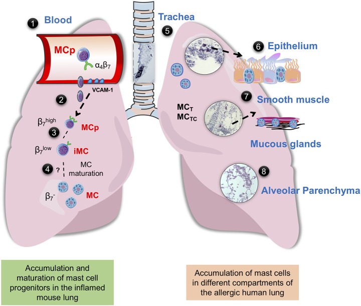
Figure 1.
Mast cells in mouse and human lung. (1) Integrin-β7+ mast cell progenitors (MCps) are found in mouse and human peripheral blood (14, 23). (2) In mice with acute allergic airway inflammation, MCps are recruited to the lungs in a process dependent on α4β1 and α4β7 integrins on the MCp and on VCAM-1 expressed in the endothelium (36). (3) After the acute phase, three mast cell (MC) populations can be identified by flow cytometry, MCps expressing high levels of integrin β7, immature/induced MCs (iMCs) expressing intermediate levels of integrin β7, and mature MCs (37, 38). (4) The iMC gradually loses the expression of integrin β7 and mature, thereby expanding the resident lung MC population. (5) In the mouse trachea and in the proximal bronchi of unprovoked mouse airways, MCs express the MC proteases mMCP-1 and-2, mMCP-4,-5-6, and-7 and CPA3, while the MCs induced by allergic airway inflammation located in the bronchovascular bundles of the lung and the epithelial lining of the large bronchi express mMCP-1,-2 and -6,-7 (29). In the human lung MCT and MCTC coexist, MCT is more frequently found in the bronchi, bronchioles and alveolar parenchyma, whereas MCTC dominates in pulmonary vessels and pleura (35) (6) In the human bronchi, patients with “Th2-high” asthma have an increased number of intraepithelial MCs (39). Genetic analyses suggest that the MCs in this location mainly express tryptase and CPA3 (39–41). (7) The number of MCs are increased in the airway smooth muscle of asthma patients (42, 43). In diseased asthma patients, there are an increased number of mast cells in the distal airway, especially in the smooth muscle and mucous glands (44). (8) Uncontrolled atopic asthmatics have a high number of mast cells in the alveolar parenchyma (45). The histology pictures shown are from hematoxylin/eosin-stained lung sections of house dust mite-sensitized wild-type BALB/c mice obtained from our unpublished experiments.

