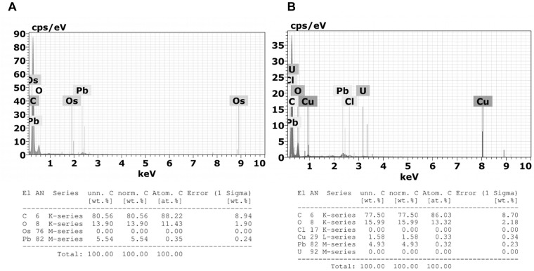
FIGURE 12.
Emission spectra obtained by Energy Dispersive X-Ray Spectroscopy (X-EDS) in TEM performed on 3 μm sections of (A) granulocytes from tween-inoculated ticks and (B) granulocytes from fungus-infected ticks. Spectra obtained focusing the incident electron beam on the granulocyte electron-dense granules of which characteristic energies of non-X-ray emission are evident. Multiple X-EDS analyses have been performed in each group; the ones that exhibited a pattern in common with the rest of the acquired spectra are shown. It is also possible to observe the presence of copper (Cu), a component of the TEM supporting grid; osmium (Os), used as fixative; lead (Pb), and uranium (U) that are used as staining.

