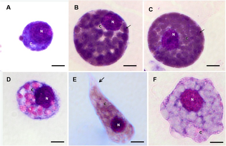
FIGURE 5.
Hemocytes representation found in the hemolymph of R. microplus engorged females under physiological conditions (untreated ticks) after Giemsa staining. (A) Prohemocyte, (B,C) Granulocyte, (D) Spherulocyte, (E) Plasmatocyte and (F) Oenocyte. Nucleus (N), cytoplasm (c), granules (thin black arrow), spherules (s) and pseudopodia (thick black arrow). Bars = 5 μm.

