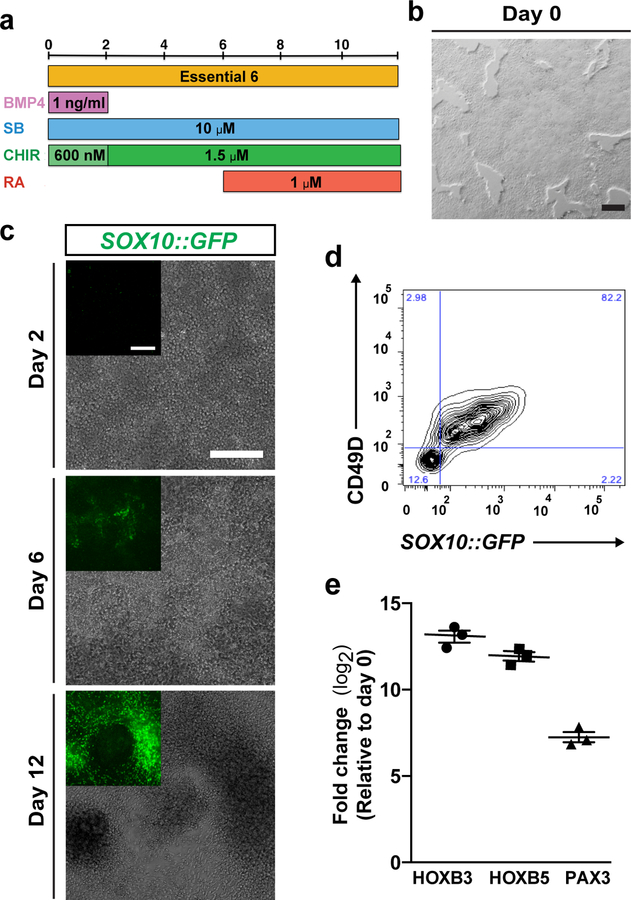
Figure 2. Induction of ENC cells from hPSCs.
a) Protocol (days 0–12) for ENC induction using option B. BMP4, Recombinant human bone morphogenetic protein-4; CHIR, CHIR 99021; RA, Retinoic Acid; SB, SB431542. b) Confluency of hPSCs on day 0 of differentiation. c) Phase contrast and SOX10::GFP reporter line GFP expression on day 2, day 6 and day 12. d) Representative image of FACS analysis of CD49D/SOX10::GFP positive ENC cells on day 12. e) Quantitative reverse transcriptase PCR (qRT–PCR) for vagal NC markers HOXB3, HOXB5, and ENC lineage marker PAX3 for ENC cells versus hPSCs. N=3 biological replicates. FC, fold change. Scale bars = 200 μm.

