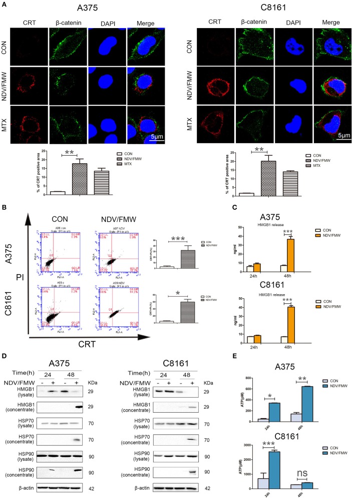
Figure 1.
NDV/FMW induces immunogenic cell death in melanoma cancer cells. (A) A375 and C8161 cells were infected with or without NDV/FMW (MOI = 1) for 48 h, to assess the translocation of calreticulin (CRT), A375 and C8161 cells were stained with an anti-CRT antibody (Red) and anti-β-catenin antibody (Green), and assessed by confocal imaging at 48 hpi of NDV/FMW (MOI = 1). β-catenin was used as a membrane marker. Mitoxantrine (MTX) was used as a positive control. DAPI was used for nuclear staining (blue). ImageJ software was used to calculate the percentage of CRT positive area (**p < 0.01). Imaging data has been quantified. Images are representative of three independent experiments. (B) A375 and C8161 cells were infected as the same in (A), the expression of CRT on the cell membrane were analyzed by flow cytometry to detect CRT in viable, PI-negative cells (*p < 0.05, ***p < 0.001). Representative dot plots (left panel) and quantification data (right panel) are shown. Data are shown for three independent replicates. (C) A375 and C8161 cells were infected with or without NDV/FMW (MOI = 1) for 24 and 48 h, release of HMGB1 in NDV/FMW-infected or mock-infected cell supernatants were detected by enzyme-linked immunosorbent (ELISA) (***p < 0.001). Data shown are representative of three independent experiments. (D) A375 and C8161 cells were infected as the same in (C), then cell lysates and the concentrated cell-free supernatants were collected. HMGB1 and HSP70/90 expression were measured by immunoblot (IB) analysis. β-actin was used as a loading control. (E) A375 and C8161 cells were infected as the same in (C), extracellular ATP was determined by ELISA (*p < 0.05). Data shown are representative of three independent experiments (n.s, not significant).

