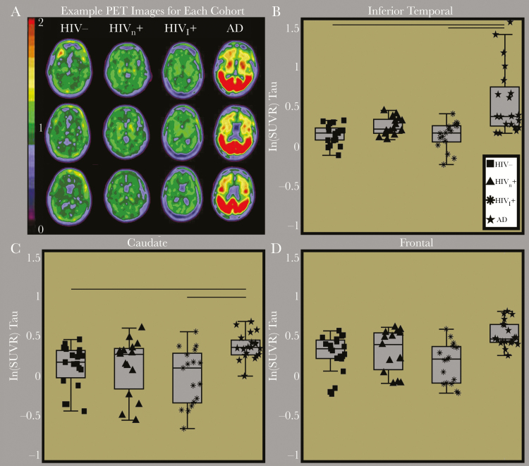
Figure 1.
A, Representative tau positron emission tomography (PET) images from a cognitively normal human immunodeficiency virus (HIV)–negative community control (HIV-), a cognitively normal HIV-infected participant (HIVn), a cognitively impaired HIV-positive participant (HIVi), and an HIV-negative participant with symptomatic Alzheimer disease (AD). On visual inspection, tau PET binding potentials were similar for the cognitively normal community control and the HIV-positive participants. Only the participant with symptomatic Alzheimer disease had significantly elevated tau PET binding. Box plots of the natural log (ln) of tau PET standardized uptake value ratios (SUVRs) for representative regions of interest, including the inferior temporal (B), caudate (C), and frontal cortex (D). Tau PET SUVRs were only elevated in participants with symptomatic Alzheimer disease, compared with the 3 other groups.

