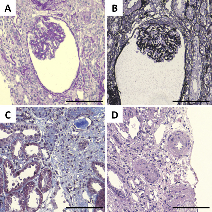
Figure 3.
Histopathology of the kidney. (A) Periodic acid-Schiff (PAS) staining showing glomerular prostration. (B) Periodic acid-methenamine-silver (PAM) staining showing basement membrane wrinkling. (C) Masson’s trichrome (MT) staining showing tubular atrophy. (D) Hematoxylin and Eosin staining showing arteriole intimal hyperplasia. Scale bars: 100 μm

