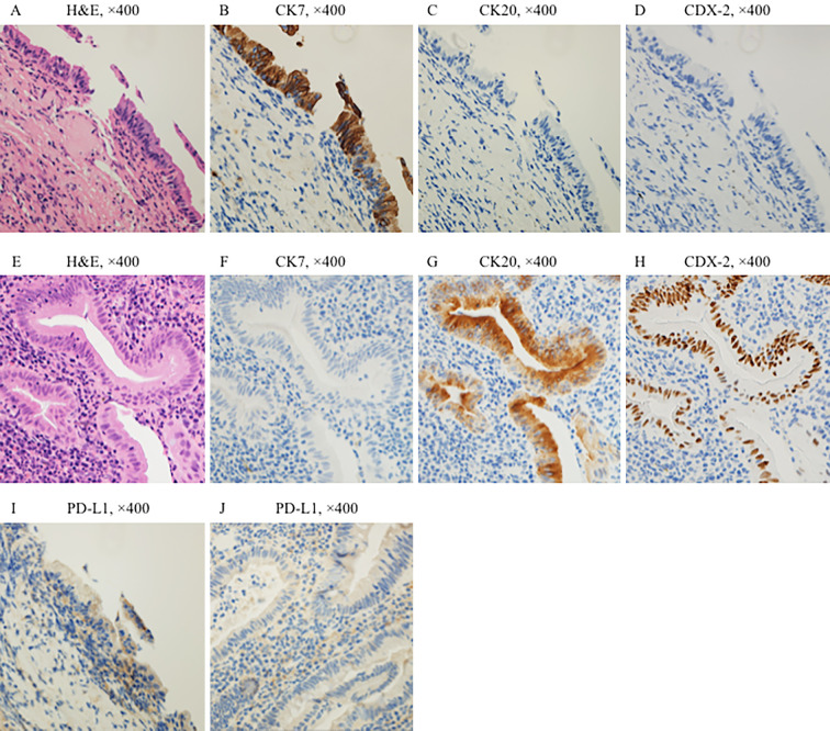
Figure 3.
Histological and immunohistochemical findings comparing lung tumor and colorectal tumor tissues. Hematoxylin and Eosin staining of the lung tumor (A) and colorectal tumor (E) are shown. Expression of cytokeratin (CK) 7, a marker of lung cancer, was observed in the tumor cells of lung biopsy tissue (B). The expression of CK 20 and caudal-type homeobox protein 2 (CDX-2), which are markers of colorectal cancer, was not observed in the tumor cells of lung biopsy tissue (C, D). In contrast, expression of CK 7 was not observed in the tumor cells of colorectal tissue (F). The expression of CK 20 and CDX-2 was observed in the tumor cells of colorectal tissue (G, H). Both lung cancer and colorectal cancer were considered as primary cancers because the IHC results were exclusive. PD-L1 expression was observed in the tumor cells of lung biopsy tissue (I). CK7 (clone OVTL12/30, DAKO), CK20 (clone IT-Ks20.8, POG), CDX-2 (clone DAK-CDX2, DAKO), PD-L1 (clone 28-8, abcam).

