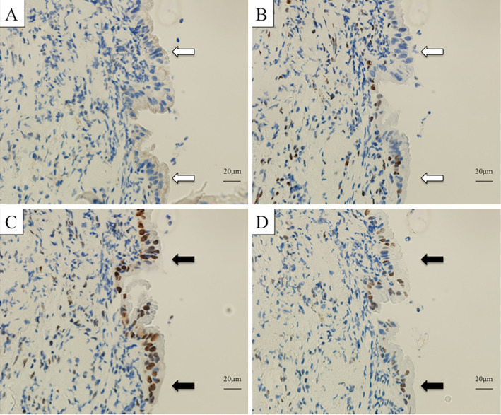
Figure 4.
A comparison of histology and immunohistochemistry (IHC) findings for lung tumor and colorectal tumor tissues. MSH2 (A) and MSH6 (B) were not expressed in the nuclei of tumor cells (white arrows), whereas MLH1 (C) and PMS2 (D) were expressed in tumor cell nuclei (black arrows). MSH2 (clone G219-1129, BD Biosciences), MSH6 (clone EPR3945, Gene Tex), MLH1 (clone ES05, Leica), PMS2 (clone A16-4, BD Pharmingen).

