Figure 3.

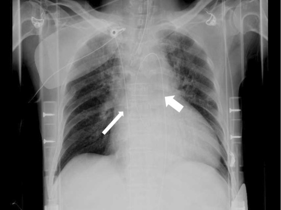
Chest X-ray after the initiation of ECLS. The thick arrow shows the wire inserted into the ascending aorta, and the thin arrow shows the wire inserted into the inferior vena cava.

Chest X-ray after the initiation of ECLS. The thick arrow shows the wire inserted into the ascending aorta, and the thin arrow shows the wire inserted into the inferior vena cava.