Figure 7.

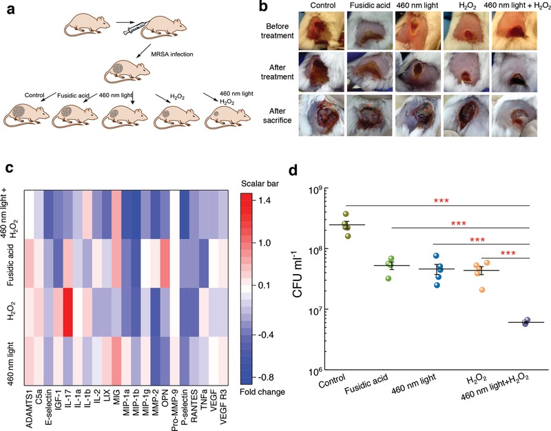
Staphyloxanthin photolysis and H2O2 effectively reduce MRSA burden in an MRSA‐infected mice wound model. a) Schematic of experiment design (not drawn to scale). b) Pictures of mice wounds of five different groups taken before treatment, after treatment, and after sacrifice. Red arrows indicate pus formation. c) Heat map of key proinflammatory cytokines and markers in the tissue homogenate samples obtained from mice treated with 460 nm light, H2O2, 460 nm light plus H2O2, or fusidic acid. Red box indicates upregulation; blue box indicates downregulation; white indicates no significant change. Scale bar represents fold change compared to the untreated group. d) MRSA CFU mL−1 after no treatment (control) or three‐consecutive‐day treatment with 2% fusidic acid (petroleum jelly as vehicle), 460 nm light, H2O2, and 460 nm light plus H2O2. Dose: 460 nm light, 24 J cm−2, H2O2, 0.045%. Error bars show the SEM from five replicates. Outlier was removed through Dixon's Q test. Unpaired two‐tailed t‐test (***: p < 0.001).
