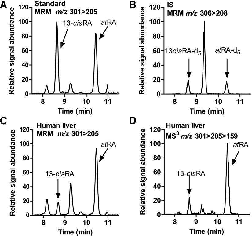
Fig. 3.
Detection of atRA and 13-cisRA in human liver. (A and B) MRM chromatograms of synthetic standards and internal standards spiked into blank serum and extracted as described in Materials and Methods. (C) MRM chromatogram of atRA and 13-cis-RA in a pooled human liver homogenate (n = 6). (D) Selected ion–extracted LC-MS/MS/MS (MS3) chromatogram (m/z 301 > 205 > 159) of atRA and 13-cisRA in the same liver sample depicted in (C).

