-
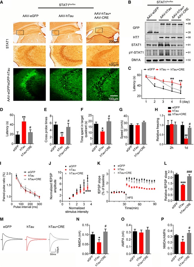
A, B
AAV‐Cre (5 × 10¹² v.g./ml) mixed with AAV‐hTau or AAV‐eGFP (1.13 × 10¹³ v.g./ml) was stereotaxically injected into the hippocampal CA3 of 3‐month‐old STAT1flox/flox mice. One month later, downregulation of STAT1 was confirmed by Western blotting and immunohistochemical staining.
-
C
Downregulation of STAT1 ameliorated hTau‐induced spatial learning deficit shown by the decreased escape latency during 5 consecutive days training in Morris water maze (MWM) test (n = 9–11 for each group).
-
D–G
Downregulation of STAT1 ameliorated hTau‐induced spatial memory deficit shown by the decreased latency to reach the platform quadrant (D), the increased crossing time in the platform site (E), and time spent in the target quadrant (F) measured at day 6 by removing the platform in MWM test; no motor dysfunction was seen (G) (n = 9–11 for each group).
-
H
Downregulation of STAT1 ameliorated hTau‐induced contextual memory deficits measured at 24 h during contextual fear conditioning test (n = 8 each group).
-
I–L
One month after the virus infection, paired‐pulse ratio (PPR) was recorded in hippocampal CA3 of hTau or STAT1 knockdown mice (I). The I/O curve of fEPSP recorded on acute hippocampal slices (J). The slope of fEPSP after HFS recorded on hippocampal slices of hTau or STAT1 knockdown mice (K). LTP magnitude was calculated as the average (normalized to baseline) of the responses recorded 40–60 min after conditioning stimulation (L).
-
M–P
One month after the virus infection, whole‐cell patch clamp was used to measure the function of NMDA (at +40 mV) and AMPA (at −70 mV) receptors on acute brain slices (400 μm). The insets show representative sample traces of EPSCs in virus‐infected neurons (M). The reduced NMDA and unchanged AMPA currents with a reduced NMDA/AMPA ratio were seen in hTau‐infected neurons, while knockdown of STAT1 restored the hTau‐induced NMDA currents (N–P). (n = 12 neurons from four animals for eGFP group; n = 11 neurons from four animals for hTau group; n = 13 neurons from four animals for hTau+CRE group).
Data information: Data were presented as mean ± SEM for (C‐H) and mean ± SD for others (two‐way repeated measures analysis of variance (ANOVA) followed by Bonferroni's post hoc test for C, two‐way analysis of variance (ANOVA) followed by Bonferroni's post hoc test for I‐K, one‐way analysis of variance (ANOVA) followed by Bonferroni's post hoc test for others). *
< 0.001 vs hTau.

