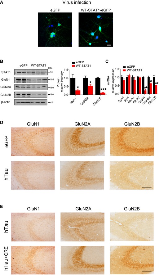
Figure EV4. STAT1 regulates the expression of NMDARs.

-
AAAV‐STAT1‐eGFP (WT‐STAT1) or the empty vector (AAV‐eGFP) (1 × 10¹² v.g./ml) was transfected into the primary hippocampal neurons (6 div) for 6 days, and expression of the virus was shown by direct fluorescence. Scale bar, 20 μm.
-
B, COverexpression of WT‐STAT1 selectively decreased protein and mRNA levels of GluN1, GluN2A, and GluN2B detected by Western blotting and qRT–PCR. *P < 0.05; **P < 0.01; ***P < 0.001 vs eGFP. Data were presented as mean ± SD (n = 4 from four independent experiments. Mann–Whitney test).
-
DAAV‐hTau‐eGFP (tau) or the empty vector AAV‐eGFP (eGFP) (1.13 × 10¹³ v.g./ml) was stereotaxically injected into hippocampal CA3 of 3‐month‐old C57 mice. After 1 month, the decreased GluN1, GluN2A, and GluN2B expression was detected by immunohistochemical staining. Scale bar, 100 μm.
-
EThe mixture of AAV‐hTau (1.13 × 10¹³ v.g./ml) and AAV‐Cre (5 × 10¹² v.g./ml) (1 μl AAV‐hTau plus 2 μl AAV‐Cre) was stereotaxically infused into the hippocampal CA3 of 3‐month‐old STAT1flox/flox mice. One month later, the restored GluN1, GluN2A, and GluN2B expression was detected by immunohistochemical staining. Scale bar, 100 μm.
Source data are available online for this figure.
