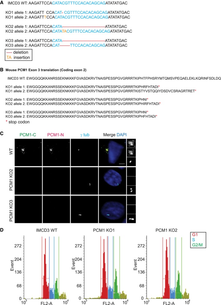
Figure EV1. IMCD3 PCM1 KO cells are devoid of satellite structures.

- IMCD3 PCM1 KO clones are all compound heterozygotes with mutations that lead to early stop codons. 1,000‐bp region around the gRNA‐target site was PCR‐amplified and cloned. Sequencing of five different clones for each line identified one‐nucleotide (nt) deletion on one allele and one‐nt insertion on the other for line 1, 16‐nt deletion on one allele and 2‐nt insertion on the other for line 2, and 16‐nt deletion for one allele and 4‐nt deletion on the other for line 3.
- Translation products on protein‐coding exon 2 of the gRNA‐targeting exon in IMCD3 KO clones.
- Immunofluorescence analysis of control and IMCD3 PCM1 KO clones. Cells were fixed and stained for centrosomes with anti‐γ‐tubulin antibody and PCM1 with PCM1‐N antibody (targeting 1–254 amino acids) and PCM1‐C antibody (targeting 1,665–2,026 amino acids). Scale bar, 4 μm.
- FACS sorting of propidium iodide‐stained IMCD3 control and PCM1 KO cells. Graphs are prepared with the cell number on y‐axis and PI fluorescence reflecting the DNA content on x‐axis.
