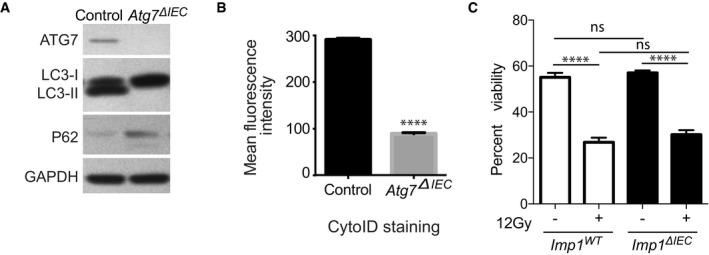
Representative western blot comparing Atg7
ΔIEC mice and controls. Atg7
ΔIEC mice show no ATG7 expression and decreased autophagic flux indicated by increased p62 and absence of LC3‐II levels.
Mean fluorescence intensity of CytoID is significantly reduced in epithelial cells From Atg7
ΔIEC mice indicating decreased autophagy (n = 3 per genotype).
Graph showing percentage of DAPI (4′,6‐diamidino‐2‐phenylindole) negative crypt cells (indicating viability) from Imp1
WT (n = 7) and Imp1
ΔIEC mice (n = 8) using flow cytometry with and without 12 Gy radiation. Although 12 Gy radiation significantly decreased viability, there was a no difference in viability between the two genotypes in either condition.
Data information: All data are expressed as mean ± SEM. ****
P < 0.0001; by two‐tailed, unpaired
t‐test (B) or 1‐way ANOVA with Bonferroni's multiple comparison test (C).

