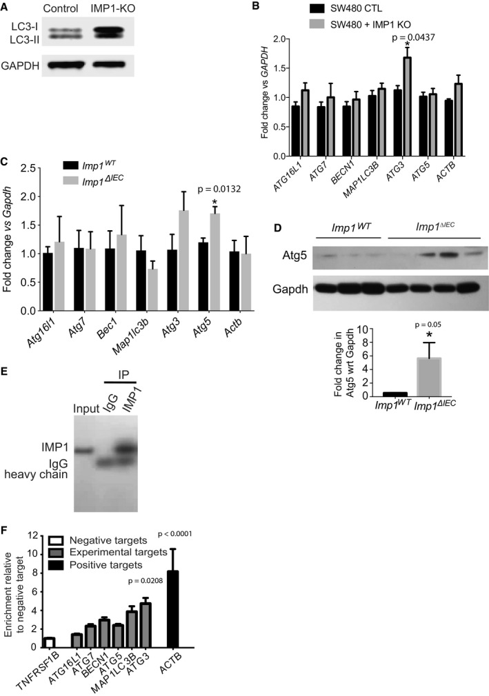
Representative western blot for LC3‐I/LC3‐II in IMP1‐KO SW480 cells.
qPCR data showing expression of different autophagy genes in SW480 cells with and without IMP1 deletion (n = 3 per genotype).
qPCR data showing expression of autophagy genes in purified epithelium from small intestinal tissue from Imp1
WT and Imp1
ΔIEC mice (n = 3 per genotype).
Immunoblot and quantification (n = 3 WT, four Imp1
ΔIEC) demonstrating upregulation of Atg5 in intestinal epithelium of Imp1
ΔIEC mice compared to controls.
RIP assay to evaluate binding of endogenous IMP1 to autophagy transcripts in Caco2 cells. Enrichment of IMP1 was confirmed by IP with either IMP1 or control IgG antibodies followed by western blot for IMP1.
Enrichment of target transcripts over control is represented relative to negative target, TNFRSF1B. Positive control was ACTB (n = 3 independent experiments).
Data information: All data are expressed as mean ± SEM. *
P < 0.05; by two‐tailed, unpaired
t‐test or ordinary 1‐way ANOVA.

