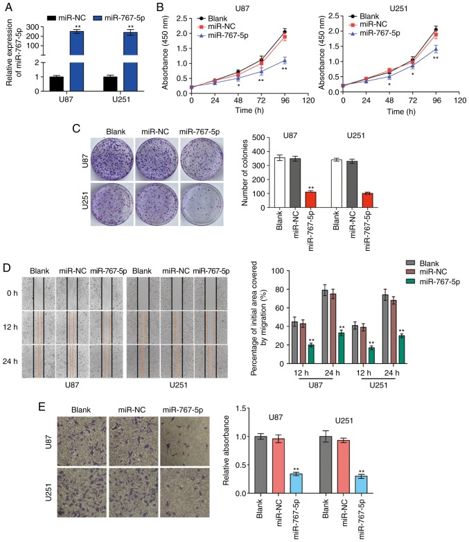
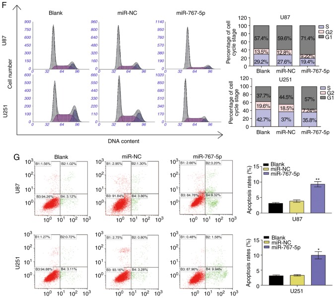
Figure 2.
Overexpression of miR-767-5p affects cancer-related behaviors of GBM cells. (A-G) For all assays, U87 and U251 cells were untransfected (blank) or transfected with a miR-767-5p mimic or a negative control sequence (miR-NC). (A) The relative expression of miR-767-5p in U87 and U251 cells was analyzed by RT-qPCR after transfection. (B) CCK-8 proliferation assay. (C) Representative images (left) and quantification (right) of colony formation assays performed after 10 days in culture. (D) Representative images (left) and quantification (right) of wound healing assays performed in U87 and U251 cells. (E) Representative images (left) and quantification (right) of Transwell migration assays. (F) Flow cytometric analysis of the cell cycle by staining with PI (DNA content). Representative histograms (left) and quantification (right) of cells in G1, G2, and S phases of the cell cycle. (G) Representative dot plots (left) and quantification (right) of an Annexin V-FITC/PI apoptosis assay performed 48 h after transfection. Data are presented as the mean ± SD of n=3. *P<0.05, **P<0.01, vs. miR-NC. GBM, glioblastoma multiforme; CCK-8, Cell Counting Kit-8; PI, propidium iodide; SD, standard deviation.


