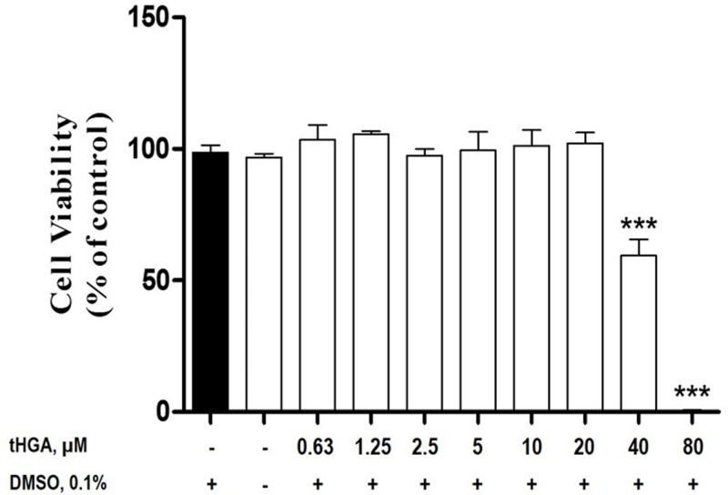
Figure 3. The cytotoxic effects of tHGA on RBL-2H3 cells.
The RBL-2H3 cells were incubated with increasing concentrations of tHGA (0-80 μM) for 24 h. The cytotoxicity of the treatments was determined by MTT assay. The results are expressed as the mean ± S.E.M values of three independent experiments. ***P<0.005 as compared with the normal RBL-2H3 cells (black bar).

