
Figure 1. Long and short isoforms of CIP4 and FBP17 have opposing effects on cortical neuronal development.
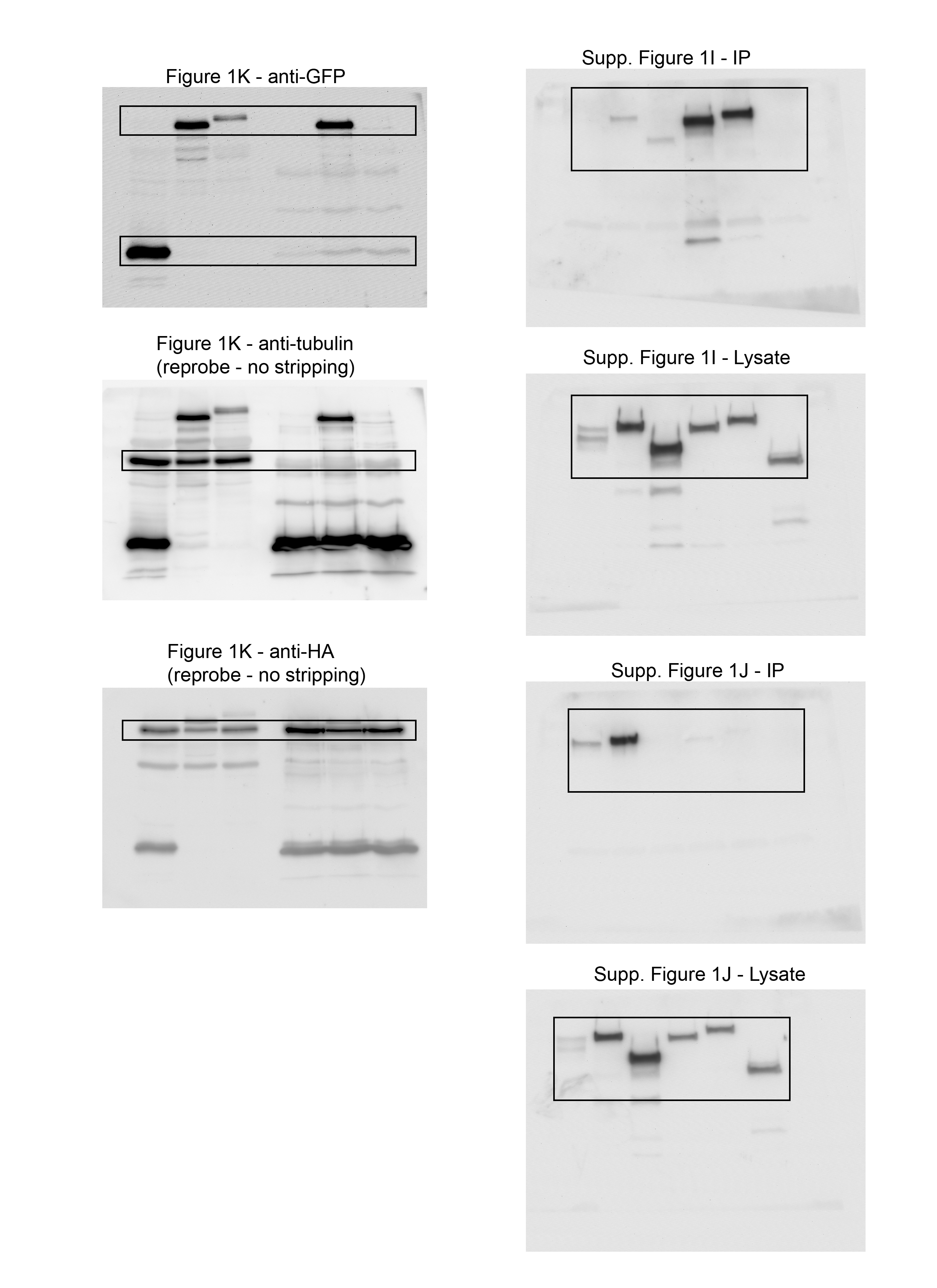
(A) Schematics of long and short human isoforms of CIP4 and FBP17. The F-BAR/EFC domain is shown as a dimer and only one C-terminal half of the protein is shown for clarity. F-BAR, HR1, and SH3 regions are false-colored, space-filling diagrams based on the following PDB files: CIP4 F-BAR/EFC domain (2EFK), FBP17 F-BAR/EFC domain (2EFL), HR1 domains (2KE4), and SH3 domains (2CT4). (B) Images of living cortical neurons at 12 h postplating, cotransfected with mRuby-Lifeact (red) to label actin and EGFP-labeled F-BAR protein (green). Contrast on black and white images is inverted for clarity. (C) Images of fixed COS-7 cells transfected with different isoforms of CIP4 and FBP17 and labeled with phalloidin (f-actin) and DAPI (nuclei). (D–G) Box-and-whisker plots showing quantification of stage 1 neurons (with points showing data that falls outside of the 10–90 percentile) comparing the effects of the different isoforms on peripheral intensity (D), filopodia number (E), cell complexity (F), and tubule number (G). CIP4S-EGFP (n = 24 cells), CIP4L-EGFP (n = 30 cells), FBP17L-EGFP (n = 23 cells), or FBP17S-EGFP (n = 31 cells). (H) Stacked bar graph comparing the percentage of neurons in stage (st.) 1, 2, and 3 for neurons expressing EGFP (n = 58), CIP4S-EGFP (n = 72), FBP17L-EGFP (n = 75), or CIP4S-tdTomato and FBP17L-EGFP (n = 65) at 12 h postplating. Two-way ANOVA with Bonferroni post-test multiple comparison. (I) Image of a living cortical neuron cotransfected with CIP4S-Scarlet and FBP17L-EGFP. (J) Box-and-whisker plot showing average colocalization (Pearson’s correlation coefficient) of CIP4S and FBP17L in cortical neurons (n = 46 cells). (K) Co-IP with CIP4S-HA and either CIP4S-EGFP or FBP17L-EGFP in cortical neurons. Original blot was separated to show higher molecular weight proteins (CIP4S-EGFP and FBP17L-EGFP) and EGFP. This blot was reprobed with antibodies to HA and tubulin. (L) Quantification of three co-IPs with CIP4S-HA. One-way ANOVA with Kruskal–Wallis post-test multiple comparisons. *P < 0.05, **P < 0.01, ***P < 0.001, and ****P < 0.0001; ns, not significant. Scale bars represent 5 µm in whole-cell images of neurons and 1 µm in insets; 15 μm in whole-cell images of COS-7 cells and 7 μm in insets.
Source data are available for this figure.
