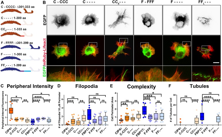
Figure 4. The F-BAR and first linker region are required for membrane binding and bending.
(A) Schematic of deletion constructs of CIP4S and FBP17L. (B) Images of living cortical neurons cotransfected with mRuby-Lifeact and EGFP-labeled protein or deletion mutant at 12 h postplating. (C–F) Quantification of stage 1 neurons comparing the effects of the deletion constructs on peripheral intensity (C), filopodia number (D), cell complexity (E), and tubule number (F) at 12 h postplating. CIP4S-EGFP (n = 24 cells), C-CCC-EGFP (n = 35 cells), C---- EGFP (n = 21 cells), CCS--- EGFP (n = 22 cells), FBP17L-EGFP (n = 23 cells), F-FFF EGFP (n = 33 cells), F---- EGFP (n = 28 cells), and FFL--- EGFP (n = 29 cells). One-way ANOVA with Kruskal–Wallis post-test multiple comparisons. *P < 0.05, **P < 0.01, ***P < 0.001, and ****P < 0.0001; ns, not significant. Scale bars represent 5 µm in whole-cell images and 1 µm in insets.

