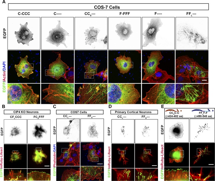
Figure S5. The L1 region is necessary for membrane tubulation in COS-7 cells and cortical neurons.
(A) Images of COS-7 cells transfected with one of the following: C-CCC-EGFP, C----EGFP, CCS---EGFP, F-FFF-EGFP, F----EGFP, or FFL---EGFP, fixed and stained with phalloidin and DAPI. (B) Images of living CIP4 KO cortical neurons transfected with mRuby-Lifeact and either CFLCCC-EGFP or FCSFFF-EGFP. (C) Images of COS-7 cells transfected with either CCL---EGFP or FFS---EGFP, fixed, and stained with phalloidin and DAPI. (D) Images of cortical neurons transfected with mRuby-Lifeact and either CCL---EGFP or FFS---EGFP. (E) Schematic representation of Linker 2 deletion constructs of CIP4S-EGFP (CCSC-C) and FBP17L-EGFP (FFLF-F). Images of living cortical neurons transfected with mRuby-Lifeact and either CCSC-C-EGFP or FFLF-F-EGFP. Scale bars represent 5 µm in whole-cell images of neurons and 1 µm in insets and 15 μm in whole-cell images of COS-7 cells and 7 μm in insets.

