Fig. 4.
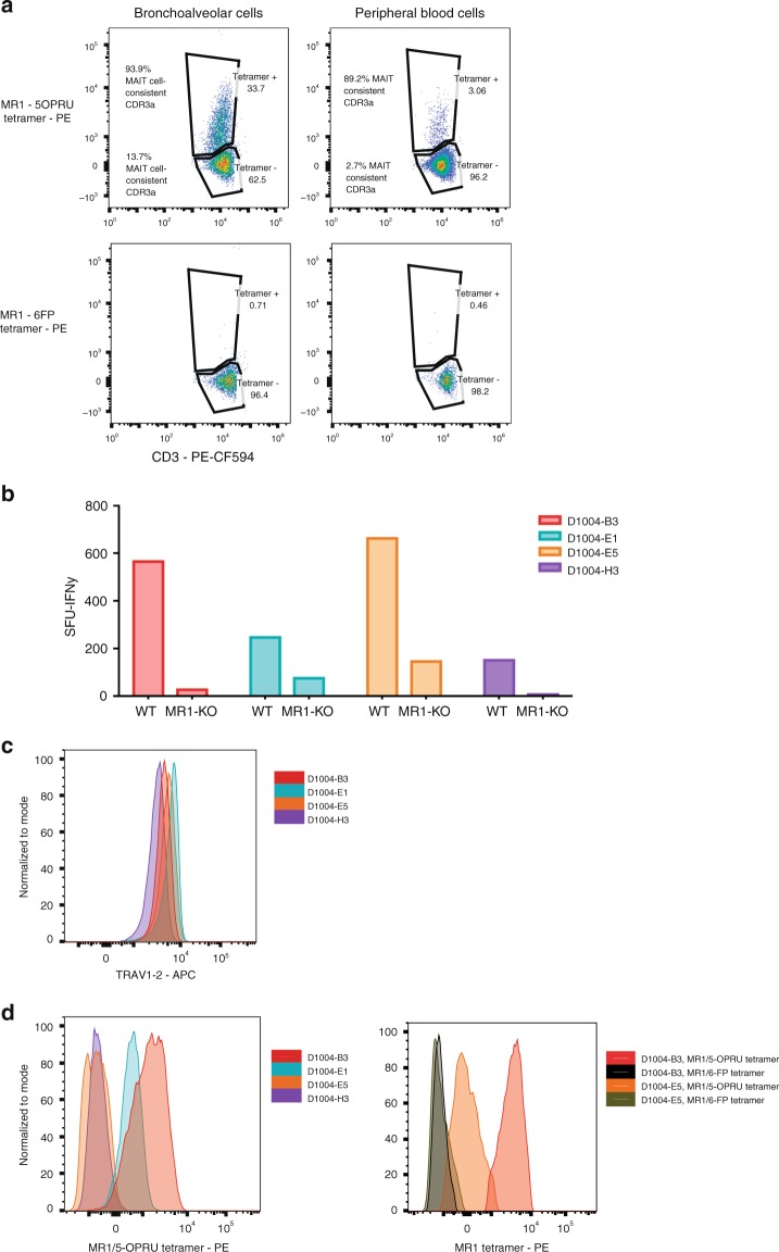
Heterogeneous MR1/5-OP-RU staining of bronchoalveolar TRAV1-2+ CD8+ T cells with MAIT cell-consistent CDR3α‘s and MR1-restricted function. a Frequency of MR1-tetramer+ cells (loaded with active (5-OP-RU) and control (6FP) ligand) in TRAV1-2+ T cells (gated on live, CD3+, CD8+ lymphocytes) from the BAL fluid and peripheral blood of a patient with TB. The proportion of cells utilizing MAIT cell-consistent CDR3α‘s (MAIT Match Score ≥.95) in MR1/5-OP-RU tetramer positive and negative populations are shown. b IFNγ spot-forming units (SFU) produced by four T cell clones generated from BAL fluid and stimulated with M. smegmatis-infected wildtype (WT) or MR1-KO A549 cells, Supplementary Data. c α-TRAV1-2 staining of four T cell clones generated from BAL fluid demonstrates consistent staining. Histograms are mode-normalized. d Binding of MR1/5-OP-RU tetramer on the same four T cell clones generated from BAL fluid demonstrates heterogenous MR1/5-OPRU tetramer staining (left). Binding of MR1/6-FP (control) and MR1/5-OPRU tetramer is shown for two clones (right). Histograms are mode-normalized

