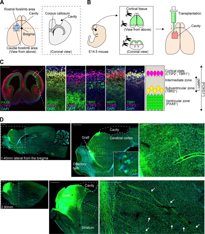
Fig. 1.
Schematic for the cortical lesions and cell transplantation, characteristics of the frontal cortex of E14.5 mice, and representative sagittal images after cell transplantation. a Schematic of the procedure for making cortical lesions. b Schematic of the cell transplantation procedure. E14.5 mouse cortical tissue is harvested and transplanted into the anterior part of the lesioned motor cortex. c Immunohistological evaluations of the anatomical cell distribution in E14.5 mouse cortex. FOXG1 is stained throughout the cortical tissues, PAX6 is stained mainly in the ventricular zone, TBR2 is stained outside of the ventricular zone, NRP1 is stained in a broad area of the tissues, including the intermediate zone, and CTIP2 and TBR1 are co-stained mainly in the cortical plate. Scale bars, 500 µm (left), 50 µm (right). Nuclear counter staining, 4,6-dimamidino-2-phenylindole (DAPI). d (Left images) Representative sagittal images of GFP staining at 1.40 mm and 2.90 mm lateral from the bregma at 2 weeks after the cell transplantation. Scale bars, 1000 µm. (Middle images) Magnified views of the white dotted frames in the left images (top), and magnified views of the white frames in the upper middle image (bottom). Scale bars, 100 µm. (Right images) Magnified views of the yellow dotted frames in the middle images. The fibers pass through the striatum (arrows). Scale bars, 50 µm. Nuclear counter staining, DAPI

