Fig. 3.
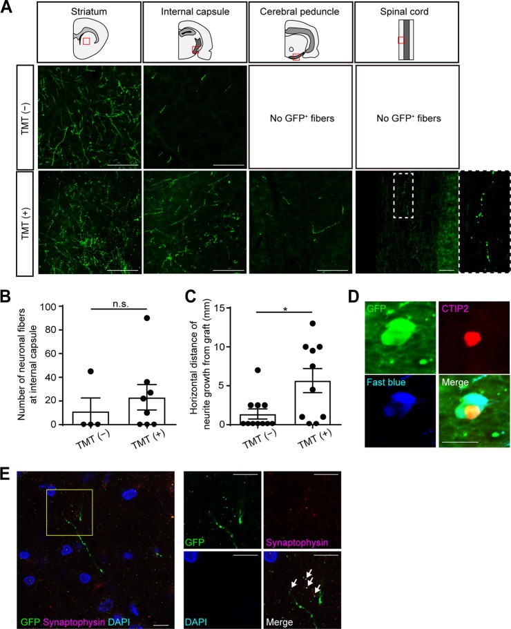
TMT promoted neurite extensions from grafted cells. a Representative images of GFP+ neurites at the ipsilateral striatum, ipsilateral internal capsule, ipsilateral cerebral peduncle, and spinal cord. Scale bars, 100 µm. In the TMT (−) group, extending GFP+ neuronal fibers were found in the ipsilateral striatum and ipsilateral internal capsule, but not in the ipsilateral cerebral peduncle or spinal cord. In the TMT (+) group, extending GFP+ neuronal fibers were found in the ipsilateral striatum, ipsilateral internal capsule, ipsilateral cerebral peduncle, and spinal cord. b Quantitative analysis of the number of GFP+ neurites at the ipsilateral internal capsule. The number of neurites was not significantly different between the TMT (−) group and the TMT (+) group. Mann–Whitney tests were performed; n = 4 in the TMT (−) group and n = 8 in the TMT (+) group. Data are presented as means ± SEM. c Quantitative analysis of the horizontal maximum distance of neuronal growth from the graft. The distance is significantly longer in the TMT (+) group than the TMT (−) group. Mann–Whitney tests were performed; p = 0.0219, n = 11 in the TMT (−) group and n = 10 in the TMT (+) group. Data are presented as means ± SEM. d Representative images of Fast blue+, GFP+ and CTIP2+ cells in the graft. In the TMT (+) group, double-labeled staining in the graft demonstrated that Fast blue+/GFP+ cells co-expressed CTIP2. Scale bar, 20 µm. e Representative images of GFP+ neurites and Synaptophysin+ vesicles on neuronal fibers around the ipsilateral red nucleus (arrows). Scale bars, 10 µm. Nuclear counter staining, 4,6-dimamidino-2-phenylindole (DAPI)

