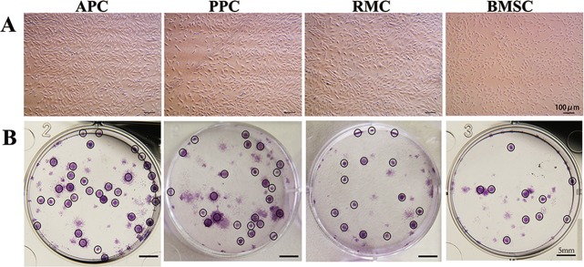
Fig. 1. Morphological observation and colony formation of AS cells.
a Cells from antler relative tissues were isolated and cultured as described in methodology section; their morphology was monitored under microscope (Passages ≤ 5); Bar = 100 μm. b Colonies formed after seeding 100 cells/well at day 14, and were stained with crystal violet dye; Bar = 5 mm; APC antlerogenic periosteal cell, PPC pedicle periosteal cell, RMC reserve mesenchymal cell, BMSC bone marrow stem cell

