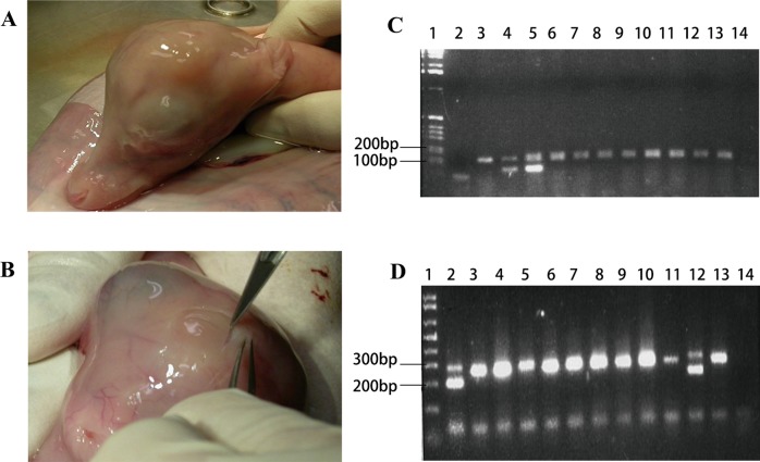
Fig. 7. Production of chimera from APCs.
a Male fetus derived from blastocyst injected with morula embryo cells; neck girth of 9.0 cm. b Female fetus derived from blastocyst injected with male AP cells. Note the prominent pedicle formation on the head; neck girth of 9.5 cm. c PCR amplification of SRY region of the Y chromosome (male 104 bp) and internal DNA positive control (194 bp) of various tissues from the female fetus with pedicles. (1) Ladder; (2) Water control; (3) Female control; (4) Male control; (5) Ovary (male DNA in the ovary); (6) Kidney; (7) Skin; (8) Pedicles; (9) Skin pedicles; (10) Heart; (11) Gut; (12) Brainstem; (13) Muscle; (14) Blood. d PCR amplification of amelogenin gene (male sequence deletion 200 bp) and female 300 bp of various tissues from the female fetus with pedicles. (1) Ladder; (2) Ovary (male DNA in the ovary); (3) Kidney; (4) Skin; (5) Pedicles; (6) Skin pedicles; (7) Heart; (8) Gut; (9) Brainstem; (10) Muscle; (11) Blood; (12) Male control; (13) Female control; (14) Water control

