Figure 6.
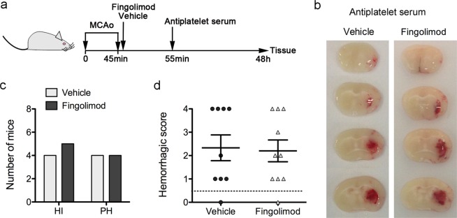
Fingolimod does not reduce hemorrhagic transformation in platelet-depleted mice after severe brain ischemia. (a) Schematic illustration of the experimental design. Immediately after reperfusion following 45-min MCAo, wild type mice received an i.p. administration of fingolimod (n = 10) (1 mg/kg) or vehicle (n = 9). Ten minutes later, all the mice received an i.p. administration of anti-platelet serum. At 48 h, fresh brain tissue was obtained to assess for the presence of blood in the ischemic tissue. (b) Representative brain sections showing hemorrhages in thrombocypenic mice regardless of whether they received vehicle or fingolimod. (c,d) A hemorrhagic score (HS) was assigned ranging from 0 (no bleeding) to 4 (large parenchymal hematoma). One mouse per group showed HS = 0. The mice showing some degree of hemorrhage were grouped according to HS categories as follows: HS = 1–2 (hemorrhagic infarction HI1 and HI2) and HS = 3–4 (parenchymal hematomas PH1 and PH2). Two of the mice in the fingolimod group with HS = 4 died at the time of sacrifice (48 h) but the brain was obtained and were included in the analysis. Fingolimod did not reduce the number of platelet-depleted mice with parenchymal hematomas (Chi-square, p = 0.819) (c), or the mean HS (Mann-Whitney test p = 0.838) (d).

