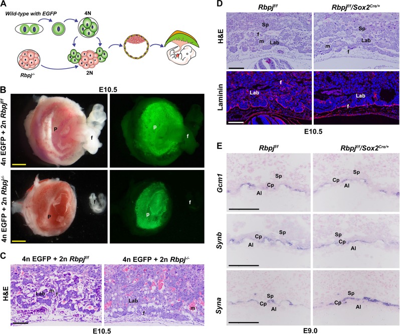
Fig. 3. Allantois-expressed Rbpj is essential for chorioallantoic fusion and branching morphogenesis.
a Diagram illustrating the procedure of tetraploid aggregation, in which one Rbpj−/− morula aggregated with two wild-type tetraploid EGFP-expressing four-cell embryos. The tetraploid cells contribute exclusively to the trophoblast cells of the placenta, rather than the allantois. b E10.5 embryos and placentas generated by tetraploid aggregation assay. c HE staining of E10.5 reconstructed placentas from tetraploid aggregation assay. d Shallow invagination of allantoic blood vessels into the chorionic ectoderm was revealed by HE and laminin staining in placentas with allantois-specific deletion of Rbpj by Sox2cre/+. Cy3-labeled Laminin is in red, DAPI-labeled nuclei in blue. e The differentiation of a chorionic trophoblast was revealed by the expressions of Gcm1 and Synb (markers for SynT-II), and Syna (marker for SynT-I). By in situ hybridization, the expression of these markers was comparable between the Rbpjf/f and Rbpjf/f/Sox2cre/+ placentas. Images in b–e are representative of at least three independent experiments. Al allantois, Cp chorion plate, Lab labyrinth, f fetal blood vessel, m maternal blood sinus, p placenta, Sp spongiotrophoblast layer. Yellow scale bars: 1 mm; white and black scale bars: 100 μm

