Fig. 5.
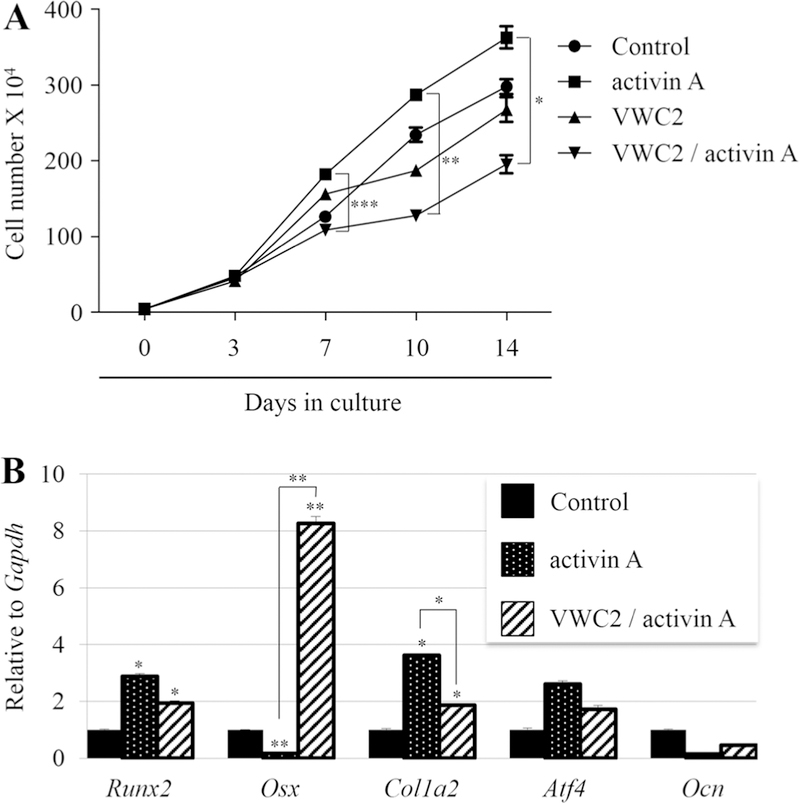
Effect of VWC2 on activin A-induced cell functions. a Effect of VWC2 on activin A-induced osteoblastic cell growth. MC3T3-E1 cells were plated in triplicate, and on the following day, cells were treated with PBS (control), activin A (250 ng/ml), VWC2 (500 ng/ml), or both VWC2 and activin A, and further cultured up to 14 days. Cell numbers were counted at each time point indicated and expressed as the mean ± SD. ***P value < 0.001, **P value < 0.01, *P value < 0.05. b Effect of VWC2 on osteoblast differentiation treated with activin A. MC3T3-E1 cells were treated with PBS (control), activin A (250 ng/ml), and VWC2 together with activin A, and further cultured for 7 days. Total RNA was extracted and the expression of osteoblastic markers was analyzed by real-time PCR. The normalized values are shown as mean + S.D. based on triplicate assays and statistically analyzed. **P value < 0.01, *P value < 0.05. Runx2; runtrelated transcription factor 2, Osx; Osterix, Col1a2; Collagen type 1 alpha 2 chain, Atf4; Activating transcription factor 4, Ocn; Osteocalcin

