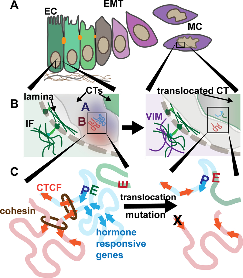
Figure 1. Cancer-compromised higher order chromatin organization.

A.) An epithelial-to-mesenchymal transition (EMT) occurs during breast cancer progression during which cells relinquish their epithelial cell (EC) tight junctions and polarity while acquiring mesenchymal cell (MC) characteristics that include migration and invasiveness. B.) An inset of a portion of the nucleus is shown. The nucleo-cytoplasmic link is illustrated wherein forces from within the cytoplasm can be transferred into the nucleus. The intermediate filament (IF) protein, vimentin (VIM), is increased in expression during EMT. Portions of two chromosome territories (CTs) are shown (grey and green). Compartments within one CT are shown. An open, euchromatic, A compartment is blue and closed, heterochromatic, B compartment is red. C) In the loop extrusion model, cohesin extrudes DNA until two convergent CTCF motifs are encountered. Genes that are responsive to a stimulus (e.g. hormones) are enriched to reside within the same TADs. Alterations in the genome that occur within cancer nuclei such as translocations, deletions, and inversions may result in the disruption of proper enhancer (E)- promoter (P) interactions and result in aberrant regulation. Mutation of CTCF binding sites are frequent in cancers and mutation of these sites has been shown to disrupt looping.
