
Figure 2. Mutations in the DNA binding- Runt homology domain of RUNX1 in breast cancer.
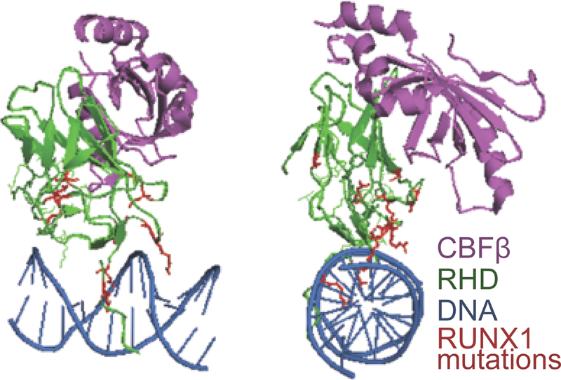
The structure of RUNX1’s Runt homology domain (RHD; rendered based on the Protein Data Bank code 1H9D293 is shown in two orientations rotated 90 degrees relative to each other (front and side). CBF‐β is shown in purple, DNA binding-RHD is in green, and DNA is in blue. Mutations found in the RHD in breast tumor patient samples (red) suggests a loss of RUNX1 function in breast cancer.