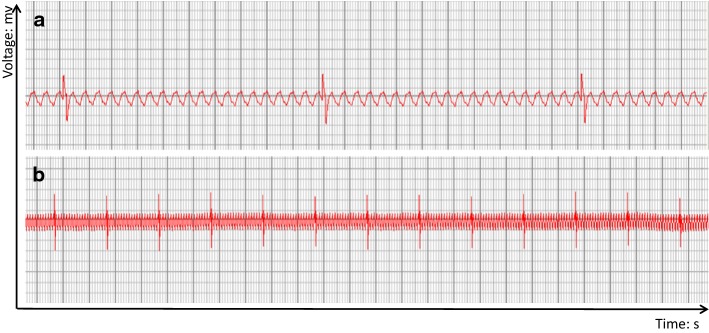
Fig. 6.
ECG of bioartificial heart. Representative images from the electrical activity assessment of the bioartificial whole heart after 10 days in a heart bioreactor. a Part of Magnified electrocardiogram of bioartificial heart. b Part of electrocardiogram of bioartificial. The abscissa is time, the smallest grid is 0.04 s, and the big grid is 0.2 s. The ordinate is mv, with 0.1 mv per small cell, thus, five small cells (one large cell) is 0.5 mv, and the length of two large cells is 1 mm

