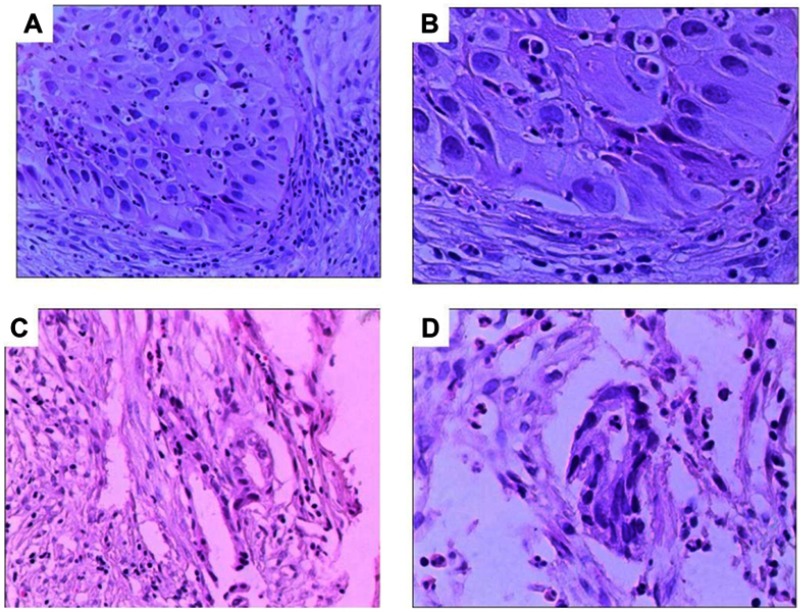
Figure 2.
Histologic features from synchronous multiple tumors. Hematoxylin and Eosin (H and E) staining revealed lung adenocarcinoma at both sides. Tissues were obtained by core needle biopsy from the bottom lobe of the right lung and the subsequent resection specimen demonstrated an Acinar-Predominant AC (A, Magnification×200 and B, Magnification×400). Tissues were obtained by core needle biopsy from the bottom lobe of the left lung and the subsequent resection specimen demonstrated an Acinar-Predominant AC (C, magnification×200 and D, magnification×400).
Abbreviation: AC, adenocarcinoma.

