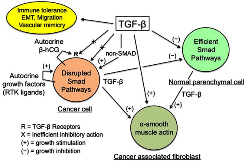
Figure 1.
TGF-β regulation of cancer tissue.
Notes: Enhanced TGF-β secretion by cancer and stromal cells promotes the development of a dense myofibroblastic microenvironment with CAFs expressing α-smooth muscle actin, together with immune tolerance and EMT, thus plagiarizing the features of the placental trophoblast-decidua interface. Conversely, inhibition of cancer cell growth by the physiological TGF-β cytostatic program is virtually nonexistent, due to various obstacles: impairment of the TGF-β/SMAD axis and of the SMAD-driven gene transcription, high expression of SMAD7, and actions of autocrine growth factors and β-hCG. The β-chain of hCG, a hormone normally produced by trophoblast, may interfere with TGF-β receptors, thus preventing the normal action of TGF-β. The above figure is valid for trophoblastic cells (in place of cancer cells), with the SMAD cytostatic program being silenced by RTK ligands and β-hCG, and for decidual myofibroblasts (in place of CAFs).

