

Abstract
Breast cancer (BC) is the most common malignancy in women worldwide, and one of the deadliest after lung cancer. Currently, standard methods for cancer therapy including BC are surgery followed by chemotherapy or radiotherapy. However, both chemotherapy and radiotherapy often fail to treat BC due to the side effects that these therapies incur in normal tissues and organs. In recent years, various nanoparticles (NPs) have been discovered and synthesized to be able to selectively target tumor cells without causing any harm to the healthy cells or organs. Therefore, NPs-mediated targeted drug delivery systems (DDS) have become a promising technique to treat BC. In addition to their selectivity to target tumor cells and reduce side effects, NPs have other unique properties which make them desirable for cancer treatment such as low toxicity, good compatibility, ease of preparation, high photoluminescence (PL) for bioimaging in vivo, and high loadability of drugs due to their tunable surface functionalities. In this study, we summarize with a critical analysis of the most recent therapeutic studies involving various NPs-mediated DDS as alternatives for the traditional treatment approaches for BC. It will shed light on the significance of NPs-mediated DDS and serve as a guide to seeking for the ideal methodology for future targeted drug delivery for an efficient BC treatment.
Keywords: Breast cancer, Drug delivery, Nanoparticles, Bioimaging, Biomarkers
Graphical abstract
1. Introduction
Cancer is a class of diseases resulting from unregulated cell growth and these abnormal cells are able to spread or invade other parts of the body. Based on the presumed origin of the tumor cells, cancers are classified into carcinoma, sarcoma, lymphoma, leukemia, germ cell tumor and blastoma. Among them, carcinoma indicates cancer that derive from the epithelial cells and it includes nearly all cancers in breast, prostate, lung, pancreas and colon [1]. Considering the damage various cancers incur, skin and lung cancers are the most common malignancies worldwide. In addition, breast cancer (BC) is the most common cancer type among women accounting for nearly 30% of all cancers [2]. In 2018, about 266,120 new cases of invasive BC have been diagnosed that will potentially cause 40,920 cases of death according to the statistics of the American Cancer Society [2]. In contrast, BC in men accounts only for 1% of all malignant breast neoplasms [3]. Also compared to women, men tend to be diagnosed for BC at an older age as 67 years [4]. Although it is the most common cancer type in women, it is considered as treatable if diagnosed at an early stage [5, 6]. However, if metastasis is achieved, it can spread through blood and lymph systems to distant organs, increasing treatment difficulties and the fatality rates rapidly.
Similar to other cancers, the traditional treatment approaches for BC include surgery, chemotherapy and radiotherapy. The primary goal of these therapies is to eradicate tumors while prolonging the survival of patients. Nonetheless, these standard methods are challenged by the advanced and metastatic tumors in terms of tumor recurrence and drug resistance. For instance, surgery is not effective in case of tumor recurrence and metastases to distant organs including bone, lung and liver. In contrast, the goal of chemotherapy is to use cytotoxic chemotherapeutic drugs either after or without surgery to interfere with tumor cell division and growth. Radiotherapy involves delivering powerful waves of energy to disrupt the tumor cell division, which results in the shrinkage or eradication of tumors. Although chemotherapy and radiotherapy are powerful cancer treatment techniques to increase the survival rate, these techniques could lead to acute and long-term adverse effects on the patients’ healthy organs [7, 8]. For instance, the chemotherapeutic agent; trastuzumab, a monoclonal antibody used to treat BC, has shown toxicity assisted with cardiac dysfunction in long-term use [9]. Furthermore, multidrug resistance is also a challenging issue caused by over-expression of certain proteins in the tumor cells. The effect of chemotherapy is often drastically reduced in this case. Radiation therapy is a local treatment that only affects the area of the tumor location. But side effects can occur due to the damages caused on the neighboring healthy tissues. Considering these adverse effects present in the classic cancer treatment approaches, novel effective alternatives need to be well sought.
Alternatively, the use of nanomaterials as an effective drug delivery method for the cancer treatment has recently gained specific interest, and ongoing investigations are aiming to optimize this method to ultimately reduce the adverse effects caused by the conventional approaches. To date, such NPs commonly used in research for drug delivery to treat BC include liposomes, mesoporous silica NPs, viral NPs, polymer-, metal- or carbon-based NPs and different drug loading techniques are used depending on the NPs such as encapsulation, covalent or electrostatic binding and adsorption. There are numerous advantages of using NPs for drug delivery: i) it solves issues related to the poor solubility and bioavailability of the drug; ii) it enhances targeted drug permeability to cancer cells and administer slow release of the drug; and iii) NPs are small (1-100 nm), nontoxic, biodegradable and highly photoluminescent particles on to which the cancer drugs can be easily loaded. The PL could endow the in vivo drug tracking ability to determine drug delivery efficacy during treatment. For the studies of targeted DDS, different types of breast tumor cell lines have been used in vitro including MDA-MB-231, MDA-MB-453, SkBr3 and MCF-7 [5, 7, 10-14]. Besides, doxorubicin (Dox) is the most popular chemotherapeutic agent for NPs mediated delivery for BC and it has also been used together with siRNAs and miRNAs in co-delivery systems. Other chemotherapeutic agents, namely paclitaxel (PTX), cisplatin, trastuzumab, fulvestrant, anastrozole, and carboplatin are also often used for phase II and III clinical trials [14-17]. Furthermore, several combinations among these therapeutics have shown synergistic effects against BC [14].
Administering drugs using a targeted DDS can help reduce the doses because the pharmacologically effective concentrations can be achieved at lower concentrations compared to untargeted administering of drugs. Side effects resulting from toxicity and damages to healthy cells and tissues could also be significantly reduced through a targeted delivery method compared to the standard chemotherapy approach. Therefore, the development of new treatment methods such as NPs-based targeted DDS as well as combination therapy has the potential to alleviate the side effects. Usage of a DDS is also of crucial importance in treatments using drug combinations or oligonucleotides, due to the needs such as delivery without premature decay and simultaneous drug administration.
2. Drugs and Breast Cancer Biomarkers
2.1. Chemotherapy drugs and side effects
Chemotherapeutic agent, Dox which is a member of the anthracycline class is heavily used in the clinical treatments for many human cancers. It is one of the most commonly used chemotherapeutic drugs for the treatment of BC either alone or in combinations with other drugs. Various studies have been conducted to understand the side effects of Dox both in vivo and clinically [18, 19]. It is well known for its high possibility in hematopoiesis and gastrointestinal or cardiac toxicity [20]. Therefore, targeted delivery can be utterly important in Dox treatments [21].
Paclitaxel (PTX) has emerged as another important and popular chemotherapeutic agent in the BC treatments. Unlike other antimicrotubulin agents, PTX promotes tubulin dimerization and inhibits microtubule depolymerization to achieve antitumor effect [22]. The lack of cross-resistance with anthracyclines is one of the major reasons for the drastic increase of intensive clinical investigation on PTX worldwide [23]. Commonly known PTX side effects are neutropenia and peripheral neuropathy [22]. Thus, PTX dose optimization and evaluation of PTX in combination therapy regimens have become a central focus in research.
Few other commonly used regimens for chemotherapy are tamoxifen, trastuzumab, cisplatin and docetaxel. Also, for adjuvant chemotherapy treatments which are used to increase the effectiveness and lower the reoccurrence, cyclophosphamide and fluorouracil are commonly administered combined with methotrexate, Dox or mitoxantrone (MTX) [24]. The mostly reported side effects on patients are fatigue, weight loss, peripheral neuropathy and nausea [25]. But several severe after effects that have been identified are heart problems, osteoporosis, lymphedema and concerns about cognitive functions [24, 26].
2.2. Biomarkers
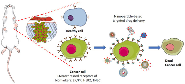
A biomarker can be described as a measurable indicator to understand biological processes or diseases from outside the patient. Currently, the clinical use of biomarkers has become inevitable in disease identification and treatments. The key aspect of the targeted nanodrug delivery in BC treatment is to target the molecular recognition markers using nanocarriers. Biomarkers targeted drug delivery improves the target specificity of drugs only towards cancer cells and less toxic to the healthy cells. Due to the overexpression of various oncogenes, biomarkers have been associated with the development and progression of resistant breast tumors. The most common BC biomarkers include estrogen receptor (ER), progesterone receptor (PR), and human epidermal growth factor receptor (HER2/ERBB2). The majority of breast tumors have ER overexpression while only approximately 25% of breast tumors have HER2 overexpression [2]. About 15% of breast tumors do not express ER, PR or HER2, classified as triple negative BC (TNBC), and are considered as the most challenging type of breast tumors [27-29]. Hence, these biomarker proteins have been used in BC classification as well as target ligands for developing novel therapeutics. Both monoclonal antibodies and anticancer drugs have been extensively tested in the treatment of BC, targeting biomarkers.
2.2.1. ER
ERs are located on the BC cell membrane as well as intracellularly. As is mentioned previously, the majority of the breast tumors are ER+ and both pre- and post-menopausal women can be affected by the ER+ type BC. Tamoxifen is the most popular antagonist for ER+ breast tumors. However, since tamoxifen does not specifically target adipose tissues, the use of NPs as nanocarrier for targeted drug delivery seems to be of significant [30]. Li et al. reported a tamoxifen delivery system based on polymer-based NPs to ER+ BC cells with a remarkably reduced cytotoxicity towards healthy cells [31]. In addition, El-Sayed and coworkers have also successfully delivered tamoxifen-conjugated NPs to ERs [32]. The system was uptaken into cancer cells mediated by the receptor, which induced the efficacy of tamoxifen up to 2.7-fold higher than the free drug.
2.2.2. PR
Similar to ER, PR is also a steroid hormone receptor and it mediates progesterone in its target tissues. It also exists in two forms, namely PR-A and PR-B [33]. PR also plays an important role in lobuloalveolar differentiation [34]. The clinical use of these is to identify the patients with invasive BC for different categories of endocrine therapy. It is used as a prediction marker for all stages of treatment such as adjuvant and neoadjuvant [35].
2.2.3. HER2
HER2 is a type of transmembrane glycogen protein that has three different regions. It has an N-terminal extracellular domain (ECD), a single α-helix transmembrane domain (TM) and an intracellular tyrosine kinase domain. HER family includes four proteins named as HER1, HER2, HER3 and HER4. HER2 is the only receptor which does not have any identified ligands, but it contributes to dimerization with the other three proteins in the family during cell growth [28]. The largest part of HER2 is the ECD which further divides into subdomains (I-IV). Cysteine rich subdomain II and IV are responsible for homodimerization and heterodimerization [28]. Subdomain II has the dimerization arm that propagates the dimerization. The monoclonal antibodies pertuzmab and trastuzumab have been identified as dimerization inhibitors. These bind to the dimerization arm of HER2 and block the signal to inhibit the dimerization with other family members retarding the cell propagation [36]. Rathinaraj et al. mentioned a covalent conjugation of trastuzumab with mercaptosuccinic acid immobilized-gold NPs which was tested with SK-BR3 BC cells [13].
2.2.4. TNBC
15% of breast tumors do not express ER, PR or HER2, which makes the targeted drug delivery more challenging. Also TNBC grow fast and aggressively making it more lethal [27]. Only 85% of TNBC are basal type but not all basal type tumors are triple negative. Basal-type tumors are similar to TNBC and they do not have ER, PR or HER2 expressions either [37]. However, some protein changes may happen to basal-type tumors, which is not common to TNBC. In many solid basal-type tumors such as BC, receptors including transferrin, folic acid, arginylglycylaspartic acid (RGD) and the epidermal growth factor receptors (EGFR) are expressed. Wu et al. introduced a NPs-mediated drug delivery system (DDS) conjugated with RGD ligand, which shows a higher cellular uptake in MDA-MB-231 BC cells than the non-targeted system [38], Also, the simultaneous delivery of a combination of therapeutic agents has shown higher success in the treatment of TNBC. The system has shown efficacy in reducing cell proliferation both in vitro and in vivo [39, 40].
3. Nanoparticles for Targeted Drug Delivery
Nanoparticles (NPs) are defined as particles (1-100 nm) with a surrounding outer layer of various organic or inorganic coatings that determine the properties of NPs. Although not frequently used in clinical treatments yet, numerous research studies are currently conducted to leverage the potential benefits of NPs in DDS for cancer therapy. NPs have been popular as nanocarriers mainly due to their characteristics such as water dispersity, biocompatibility and biodegradability. Use of NPs in cancer treatment increases the solubility and half-life of drugs thus, increasing the bioavailability of many chemotherapeutic drugs [41-43]. Also, NPs can increase the drug accumulation in the cancer tissues via the enhanced permeability and retention (EPR) [44]. Ultimately, NPs-anti-cancer drug combination can improve the efficiency of the therapy by reducing the side effects through targeting specific cancer sites using target ligands [45-47]. There are various types of NPs that have been used for BC targeted DDS. These NPs can be categorized into liposomal, polymer-, metal-, carbon-, protein-based and mesoporous silica NPs as shown in Fig 1. In addition, Table 1 summarizes some properties of these NPs in BC treatment.
Figure 1.

A diagram of different types of NPs used in BC research for targeted DDS.
Table 1.
Some of the NPs and their properties used for BC treatment.
| NPs Type | Solubility | Partic le Size (nm) |
Synthesis | Popular drugs /genes |
Cell lines |
Targeted biomarkers/recep tors |
Referen ce |
|---|---|---|---|---|---|---|---|
| Liposomal NPs | amphiphilic | 100 - 300 | Hydrophili c inner core and hydrophobi c lipid bilayer | DOX, PTX, siRNA, mRNA, siHIF1-α, siVEGF | MDA-MB-231 MDA-MB-435 MCF-7 JIMT-1 | HER2 | 48-59 |
| Polymer-based NPs | Highly hydrophilic and permeable | 100-300 | Natural polymers including cellulose and chitosan; Synthetic polymers including PVA, PEG, PLGA | DOX, PTX, Trastuzumab, Cis-platin, siRNA | MDA-MB-231 | HER2 TNBC | 15, 63-69 |
| Gold NPs | Hydrophilic | 10-20 | Mostly made up of Au3+ reduction by citrate and organic surface coating is useful for target the biomarkers | Dox, MTX, SMTX | MDA-MB-231 MCF-7 MCF-10 | EGFR, VEGFR2 | 85-89 |
| Superparamagn etic Iron Oxide NPs | N/A | 1-100 | Magnetic inner core which is made up of magnetite, Fe3O4, or maghemite, γ-Fe2O3 | DOX | MDA-MB-231 MCF7 | HER2 | 97-102 |
| Quantum Dots | Resuspendable | 2-10 | Made up of heavy metals such as Cd,In, Se,Te | Monoclonal antibodies | MDA-MB-231 MCF7 BT474 | ER, PR, HER2, EGFR | 106-111 |
| Mesoporous silica NPs | N/A | N/A | Inorganic nanomaterial which has large surface area and variable pore sizes | DOX, siRNA | MCF7 BT474 | HER2 | 117, 119, 120 |
| Carbon nanotubes | Low solubility | 1-100 | Cylindrical shape single wall or Multi wall nanomaterials which are allotropes of fullerenes. | DOX PTX | SK-BR-3 MCF7 | HER2 | 126-143 |
| Carbon dots | Resuspendable | 1-10 | Spherical shape nanoparticles with variable surface functionality | N/A | MCF-7 MDA-MB-231 | N/A | 11,158-165 |
| Protein based viral NPs | N/A | N/A | Protein envelops or capsids of viruses | DOX Trastuzumab | MCF7 MDA-MB-231 | HER2 | 163-172 |
3.1. Liposomal NPs
Liposomal NPs (LNPs) are spherical vesicles which are formulated through incorporating one or more phospholipid bilayers and their size can reach up to a few hundred nanometers. The LNPs contain a hydrophilic inner core which is surrounded by the hydrophobic lipid bilayer. Because of this unique morphology, usually hydrophobic therapeutic agents are encapsulated in the phospholipid bilayer for delivery. LNPs are also popular therapeutic carriers for hydrophilic agents by encapsulating in the inner core. Drug encapsulation also helps to drastically reduce the toxicity of drugs due to non-target distribution. Furthermore, amphiphilic agents can also be encapsulated into the aqueous inner core of the LNPs, such as vincristine and Dox [48], which specifically has been found to lower the cardio-cytotoxicity of Dox than its unencapsulated form [49]. In a study of PTX encapsulation and biological response using LNPs, Marcial et al. demonstrated that PTX encapsulated in nanostructured lipid carriers (size-75 nm) were significantly effective against MCF-7 (half maximal inhibitory concentration, IC50, −25.33±3.17 nm) and MDA-MB-231 (IC50 −2.13±0.21 nM) BC cells whereas the IC50 of free drug exceeded 500 nM [50].
LNPs tend to accumulate in tumor cells by incorporating the bilayer through the cell membrane. It has been reported that by surface modification of LNPs using PEG, longer halflives could be achieved, and the targeting efficacy also increases [51]. PEGylated LNPs showed effective targeting through passive strategies both in vitro and in vivo. LNPs have also been used to deliver combinations of drugs to achieve synergic effects by encapsulating more than one therapeutic agent in the LNPs. Wong and Chiu introduced a co-encapsulation method of both vincristine and quercetin in a PEGlyated liposome for the treatment of hormone- and trastuzumab-insensitive BC. This study showed that the co-encapsulation achieved higher synergism, prolonged drug circulation in plasma and controlled release in vivo for JIMT-1 cells. Moreover, the liposomal encapsulation has proved to be the most effective approach for the growth inhibition of JIMT-1 cells in comparison to the two individual drugs [52]. A non-PEGlyated LNPs system has also been fabricated to deliver a combination of Dox and cyclophosphamide for the treatment of metastatic BC [53].
LNPs have been identified as an effective delivery model for oligonucleotides, peptides and siRNA-based gene therapy for BC. The use of LNPs to encapsulate these peptides and nucleotides prevents their degradation in vasculature environment and allow targeted delivery by using target ligands [54, 55]. Cao et al. showed a design of surface modified LNPs with A7R-cystein peptide for PTX delivery to MDA-MB-231 cells in vitro and in vivo [56]. The study showed that A7R-cystein peptide enhanced the vesicle uptake thus increasing the cytotoxicity and accumulation in the BC xenografts, which confirmed the importance of peptide as targeting ligand in the PTX targeted delivery. Another study introduced a co-delivery system of siRNA in vitro using chitosan-coated LNPs. In this study siHIF1-α (hypoxia inducible factors) and siVEGF (vascular endothelial growth factors) were co-delivered to achieve lower cytotoxicity and higher silencing efficiency. The expressions of respective mRNAs significantly suppressed the growth of MCF-7 and MDA-MB-435 BC cells. They also explained that the chitosan-coated LNPs enhanced the stability of siRNAs by protecting them from serum degradation even after 24 h of incubation [57]. A carrier combination of bio-nanocapsule that derived from the antigen on the surface of hepatitis B virus and liposomes has also been reported for the delivery of siRNA to HER2-expressing BC. The gene silencing and protein knockdown were achieved through this system [58]. Chen et al. reported a cationic and anionic liposomal technique to overcome multidrug resistance (MDR) of BC by using Dox along with siRNA [59]. This study exhibited the fabrication of liposome-polycation-DNA (LPD) NPs using a guanidinium-containing cationic lipid which induces reactive oxygen species (ROS) and downregulates the MDR expression. Increased Dox cell uptake was observed when combined with siVEGF and targeted passive metastatic BC. Higher entrapment efficiency of Dox was observed in anionic-LPD NPs, which was modified to overcome Pgp-mediated drug efflux. Therefore, LNPs have been very popular as a nanocarrier for easily biodegradable therapeutic agents since these therapeutics can be encapsulated and protected until they reach the target cells, which is especially important for peptides and siRNAs. Although amphiphilic therapeutics can be encapsulated in LNPs the size of the LNPs is considerably larger (<50 nm), which could pose a disadvantage for nanodelivery. Moreover, to achieve better biocompatibility LNPs are often coated with polymers which increases the size further. The drug release process by opening the phospholipid bilayer could also be further discussed.
3.2. Polymer-based NPs
Polymer-based NPs (PNPs) are colloidal particles in the size order of a few hundred nanometers. These NPs are usually formulated by binding a copolymer to another polymer matrix. Polymers used in this regard can be natural products such as cellulose and chitosan [60]. On the other hand, synthetic polymers can also be used to prepare PNPs to achieve specific chemical and biological functions which make these PNPs highly demanding for biomedical applications [61]. Nanoprecipitation, emulsification and salting-out are common methodologies for chemical synthesis of PNPs [62]. These chemical syntheses of PNPs can be designed to have the required lipophilicity, charge and biocompatibility to suit a certain drug to be carried to the specific target [63]. The anti-cancer drug can be loaded onto the surface of PNPs by surface adsorption, chemical conjugation or by encapsulating into the PNPs-based DDS to be delivered to the target site [64]. Most PNPs have high solubility and permeability enabling them to be stable with slow drug release over a long period of time making these PNPs efficient nanocarriers for less hydrophilic anti-cancer agents. Moreover, it has been found that PNPs have high drug loadability and low toxicity especially when they were coated with a PEG-phospholipid copolymer [65]. Numerous clinically used chemotherapeutic agents such as Dox, PTX, trastuzumab and cis-platin have been tested for PNPs-drug conjugation. Various PNPs including polyhydroxyalkanoates, PLGA, cyclodextrins-derived PNPs have been studied as nanocarriers for various cancer treatments [63]. PEG-modified poly(ε-caprolactone) PNPs were reported for targeted delivery of tamoxifen in BC [66]. This study showed significant increase of drug accumulation in target BC cells by using the PEG-modified NPs than the unmodified NPs. A combination of N-(2-hydroxypropyl) methacrylamide with a tyrosine kinase inhibitor was also studied as a HER2-targeted DDS for HER2-overexpressing metastatic BC [67].
Moreover, different approaches of cancer inhibition have been researched apart from the use of traditional chemotherapeutic agents. Jin et al. recently reported a photodynamic therapy based on conjugated PNPs for TNBC treatment [68]. Their study showed that the luminescent conjugated PNPs could produce ROS upon light irradiation. Their synthesis of a cyclic arginine-glycine-aspartic acid peptide-decorated conjugated PNPs with poly [2-methoxy-5-(2-ethyl-hexyloxy)-1,4-phenylenevinylene] as photosensitizer had negligible cytotoxicity and could selectively kill the αvβ3 integrin-overexpressing MDA-MB-231 cells, a subtype of TNBC cells. Another study exhibited an enhanced cleavage of DNA of tumor cells, which was achieved through a tamoxifen-embedded PLGA-derived PNPs system [69]. The PLGA-tamoxifen NPs were synthesized using an emulsified nanoprecipitation technique. These PLGA-tamoxifen NPs showed higher nuclear fragmentation, cytotoxicity and greater bioavailability compared to pure tamoxifen via receptor-mediated endocytosis in both Dalton’s lymphoma ascites and MDA-MB-231 BC cells. Another different synthesis approach, a controlled layer-by-layer NPs fabrication process was developed by Deng et al. for the co-delivery of siRNA and Dox for a TNBC treatment [15] (Fig. 2). They have synthesized the PNPs using poly-L-arginine (PLA) and have noticed the size of PNPs increased up to 140 nm with the increment of layers. They concluded that the layer-by-layer technique could have high loading capability of chemotherapeutic agents. And the combination of siRNA and Dox, which targeted the multidrug resistance protein 1 (MRP1) drug efflux pump and TNBC in animal models, respectively, has shown a significantly high drug efficacy. Thus, PNPs-based DDS is another approach besides the use of LNPs. Although, the different synthetic approaches allow the PNPs to achieve certain desired characteristics, the sizes of these PNPs still are in range of about few hundred nanometers, which renders particles larger than even the LNPs, thus affecting the biodistribution [70]. Furthermore, the enhanced stability of PNPs could be disadvantages in terms of biodegradability and accumulation in the reticular-endothelial system.
Figure 2.

The schematic illustration of the layer-by-layer drug delivery platform of poly-L-arginine NPs. The figure is adapted with permission from ref. [15].
In addition, nanofibers are another group of polymeric NPs as potential candidate for drug delivery applications as well as numerous others such as cosmetics, filtration, sensors, nanoelectronics. Nanofibers manifest excellent surface area to volume ratio (approximately 100 times larger than that of microfibers), flexibility and mechanical properties such as tensile strength. As for the starting materials, polymers including polyvinylalanine (PVA), PLA, PEG and chitosan have all been used for the production of nanofibers for drug delivery applications [71]. Regarding synthetic approaches, such materials can be fabricated through drawing, template synthesis, self-assembly, phase separation and electrospinning [72]. For instance, Jayakumar et al. utilized electrospinning, which was based on the deposition of a polymer compound onto a collector under the presence of an electric field, to fabricate chitin and chitosan nanofibers [73]. On the other hand, Marty et al. fabricated nanofiber DDS to assess the cell movement in patients with metastatic BC cells [74]. Although nanofibers could provide a new approach for DDS, the toxicity and the drug loadability are concerns for major drawbacks [75].
3.3. Metal-based NPs
The metal-based NPs can be categorized also as inorganic NPs. The capabilities of these inorganic NPs have also been extensively investigated over the past decade owing to their therapeutic and imaging properties. Most of these NPs share the same type of structure containing a core which is responsible for the electronic, magnetic and optical properties and a shell which is mainly an organic surface coating. Among them, most widely used metal-based NPs for BC treatment are gold NPs, superparamagnetic iron oxide NPs (SPIONs) and quantum dots (QDs).
3.3.1. Gold NPs
Gold NPs (AuNPs) have been synthesized during the past few decades for various applications by tailoring their size [76], shape [77] and surface functionalities [78]. The most common synthesis of AuNPs involves the Au3+ reduction by citrate in aqueous media. These AuNPs have been used extensively in DDS due to the controllable unique properties and significantly low cytotoxicity as reported [79-83]. The organic surface coating is important for the DDS for targeting specific receptors/biomarkers. Thiolates and disulfides are widely used for these surface coatings mainly due to their affinity to bind onto the surface of Au. Afterwards, the drugs or therapeutic agents can be loaded on AuNPs by binding onto the surface through covalent or non-covalent bonds. The drug release could be administered at the target site depending on the loading method [79, 84], Fig. 3 is a graphic illustration of the potential Dox release triggered by pH considering the Dox conjugation with AuNPs via a pH-sensitive hydrazine band. Jafarizad et al. introduced the use of reduced graphene oxide-gold NPs for the drug delivery of covalently bonded drugs to the BC cells [85]. In this study, anticancer drug, MTX was used. The MTX was first covalently linked to 3-mercaptopropionic acid to synthesize MTX terminated thiol molecules, which were then used to functionalize the AuNPs. The formation of nanocomposite was achieved by mixing the functionalized AuNPs in a reduced graphene oxide (RGO) dispersion. It showed a DLS hydrodynamic size of the AuNPs before thiol group functionalization was with a mean diameter of 7.1 nm while, on the other hand, the hydrodynamic size after the thiol MTX (SMTX) functionalization onto the AuNPs increased up to a mean diameter of 14.9 nm confirming the functionalization. A comparison of drug release ability triggered by pH changes between the SMTX-AuNPs and SMTX-AuNPs/RGO shows that the drug release from the nanocomposite of AuNPs/RGO is significantly lower than the AuNPs. This effect could be from the slow hydrolysis of the amide bond or due to the π-stacking barrier of the reduced graphene oxide sheets. The in vitro studies have been conducted on MCF-7 BC cells.
Figure 3.

Schematic illustration of the Au-Poly(L-aspartate-DOX)-b-PEG-OH/*FA NP and its pH-triggered drug release (*FA – Folic acid). The figure is adapted with permission from ref.[84].
Receptor targeting using transferrin has been used in several different cancer studies owing to the fact that cancer cell membranes contain high amount of transferrin receptors compared to that of healthy cells. Simultaneously, several studies on BC have also been performed to target transferrin receptors on BC cells. By conjugating transferrin to AuNPs, the cellular uptake of AuNPs by BC cells can be increased for the targeted therapy [86]. Another study showed a significant inhibition of BC cells by targeting EGFR/VEGFR-2 that plays a major role in metastasis BC by increasing cell proliferation and angiogenesis. This study reported that AuNPs loaded with quercetin could inhibit the epithelial-mesenchymal transition which contributes to BC malignancies in both MCF-7 and MDA-MB-231 cells. It has shown significant decreases in several protein expressions in response to this DDS such as vimentin, N-cadherin, MMP-9, p-EGFR, VEGFR-2, p-PI3K, Akt and p-GSK3β while the DDS helped in the enhancement of E-cadherin protein expression [87]. The functionalization of AuNPs with various ligands or functional molecules helps stabilize the AuNPs significantly compared to the naked AuNPs and also enhances the cancer killing or protein expression inhibitions. In another study, AuNPs have been used to enhance the radiotherapy activity to kill BC cells. This study involved both MCF-7 and non-malignant MCF-10A cell lines. AuNPs decorated with two different functionalization molecules, cysteamine and thioglucose, have been used for the treatment. And they illustrated that although the cytoplasm distribution of these two functionalized AuNPs differed from each other, both molecules enhanced the effectiveness of the radiotherapy treatment in cancer killing compared to the non-AuNPs treated cells [88]. Another investigation showed that the Dox loaded AuNPs were able to overcome the MDR in MCF-7/ADR cancer cells [89]. AuNPs have been used in drug carrier studies extensively due to their unique characteristics especially including easy imaging through microscopic techniques such as transition electron microscopy (TEM) and controllable functionalization. One major drawback of the AuNPs could be their increased stability against the biodegradability in a biological system despite the reported low cytotoxicity.
3.3.2. Superparamagnetic Iron Oxide NPs
Superparamagnetic Iron Oxide NPs (SPIONs) are NPs of size in between 1–100 nm. These NPs have a magnetic inner core which is comprised of magnetite, Fe3O4, or maghemite, γ-Fe2O3. Maghemite is considered as one of the most suitable inner core materials for SPIONs due to its least likely toxicity from Fe(III) in the body unlike Fe(II) released from magnetite [90-93]. Direct use of SPIONs in therapeutic and biomedical applications can result in biofouling and agglomeration of these NPs in blood plasma, which is a huge drawback of direct use [94]. Thus, this magnetic core is covered by a hydrophilic coating for stabilization such as polymers which allow targeted delivery of biomolecules to specific sites. Most commonly used stabilization biopolymers include polysaccharides, PEG, dextran, alginate and polyacrylic acid [95, 96]. SPIONs have received increased popularity for immunoassays, tissue repairs, chemotherapy and magnetic resonance imaging (MRI) as contrast agents due to the inherent properties such as great biocompatibility and magnetism through which therapeutics can be guided to target site using external magnetic fields and magnetic visualization can be achieved. Marcu et al. reported a SPIONs synthesis through laser pyrolysis with uniform size of about 8-10 nm in diameter [97]. They observed an effective antitumor activity of SPIONs on MCF-7 cells after further coating the NPs with antracyclinic antibiotic violamycine B1. Poller et al. also compared the impact on BC cells of three different types of SPIONs that varied in size, shape, zeta potential and surface coating. Their effects on cellular uptake, magnetic properties and cytotoxicity of SPIONs were studied in comparison [98]. From the three types of SPIONs that were used for BC treatment including dextran-coated (SPIONDEX), lauric acid coated (SPIONLA) and SPIONLA with the addition of human serum albumin (SPIONLA-HSA), SPIONLA showed the highest cellular uptake and the cell cytotoxicity towards the BC cells.
MRI is one of the frontier noninvasive visualization methods to identify tumors and other relevant targets in biomedical imaging and clinical diagnosis. An MRI measurement of tyrosine kinase HER2/neu receptor in BC cells using SPIONs was reported. Streptavidin-conjugated SPIONs were used as the targeted MRI contrast agent in this study. These NPs were directed to a panel of MCF-7 BC cells containing different amounts of the receptors which were pre-labeled with a biotinylated monoclonal antibody. The contrast of the observed imaging was illustrated to be proportional to the expression level of HER2/neu receptors. Also, an interesting observation was that the SPIONs were able to only attach to the surface of the BC cells without entering intracellularly, which is an advantage in the in vivo imaging [99]. Thus, SPIONs are capable of potentially enhancing the contrast in imaging compared to the conventional techniques [100].
Another interesting technique of reducing the size of cancers is by involving heat. Tumor cells are susceptible to heat and can be killed via treatments such as magnetic hyperthermia. But hypothermia alone is not an effective method of tumor reduction. It is often used in combination with other techniques as radiotherapy or chemotherapy for clinical use [101]. However, SPIONs functionalized electrostatically with either Nucant multivalent pseudopeptide (N6L) (MF66-N6L), Dox (MF66-DOX) or both (MF66-N6L-DOX) act as potential antitumor therapeutics on MDA-MB-231 BC bearing female athymic nude mice exposed to an altered magnetic field. The therapeutic cytotoxicity effect of magnetic hypothermia has been reported to increase while the tumor volume decreased in the combination functionalization of both N6L and DOX [102] (Fig. 4). Thus, SPIONs can be synthesized with control in size, shape and mainly surface functionalization to suit different applications.
Figure 4.

Individual temperature dosages over tumor areas. (a). By using tumor surface temperature during hyperthermia treatment, median temperature dosages were calculated as cumulative equivalent minutes (CEM43T90) and displayed as box plots. (b). Example of a treatment sequence within the alternating magnetic field (AMF), the corresponding temperature distribution over the tumor surface and the effect on tumor volume. (c). Intratumoral distribution of SPIONs (MF66-N6LDOX) was determined using micro computed tomography 24 h prior to the first hyperthermia treatment. The figure is adapted with permission from ref. [102].
3.3.3. Quantum Dots
Imaging of cancer cells is essential to track cancer progression and drug efficacy during treatment. Thus, quantum dots (QDs) play an important role owing to its excellent optical properties in the long-run of tumors imaging [103, 104]. QDs are semiconductor nanocrystals with particle size ranging between 2-10 nm. These NPs usually are comprised of a metal inner core which exhibits a narrow emission spectrum with a size-dependent emission ranging from visible to infrared (IR) light. The shell could comprise of doped metals or semiconductor layers varying according to the applications. Conjugation of QDs with surface modifing ligands and peptides allows them to be used in target-specific cancer studies. Compared with most NPs mentioned previously, significantly, QDs enabled in vivo cellular imaging due to their excellent tunable optical properties, large surface to volume ratio, high brightness and resistance towards photobleaching. However, one drawback of these QDs is their high hydrophobicity. Thus, they require surface coating with polymers or by multilayer ligand shells to gain certain water solubility [105]. Conjugation with PEG reduces the potential toxic deposition of QDs in the reticular-endothelial system and allows better surface decoration [106-108]. QDs-based multiplexed imaging has been performed for in situ better than conventional methods which obtain biomarker information at a time such as immunofluorescence and western blot [109] (Fig. 5).
Figure 5.

Optical properties and potential applications of QDs in BC research studies. Commonly used QDs are core–shell structure encapsulated with amphiphilic polymers carrying chemically active groups. Compared with traditional organic dyes, QDs show excellent optical properties (A). After being coupled with active molecules, QDs can be adapted for tissues imaging, such as studying biomarker interactions (B) and evaluating prognostic biomarkers (C), and for in vivo imaging such as mapping auxiliary lymphatic system (D), showing BC (BC) xenograft (E) and detecting BC metastasis (F) in BC research studies. The figure is adapted with permission from ref. [109]
In a study of MCF-7 and BT-474 cell lines, QDs that could emit at multiple wavelengths were introduced. These two cell lines were found to express different levels of the following five biomarkers, ER, PR, EGFR, mTOR and HER2. Thus, the QDs were conjugated with the primary antibodies of these protein biomarkers and used for simultaneous quantitative and multicolor detection of those biomarkers [110], Sun et al. mentioned a type of CuInS2/ZnS QDs-based water-soluble imaging agent for the detection of BC cells after conjugating with anti-Ki-67 monoclonal antibody, a nuclear protein associated with the cell cycle, Ki-67 [111]. The hydrophobic QDs were coated with octadecylamine and then encapsulated in an amphiphilic polymer before conjugating with the monoclonal antibody. They revealed that the optical properties of the naked QDs remained unchanged in the decorated probe and no distinct toxicity was observed in vitro in MDA-MB-231 BC cells. But slight changes were observed in the cell shapes and the overall viability of the cells. The major drawback of QDs is that its inner core is commonly comprised of heavy metals which could be toxic to human body in the long-run through accumulation in organs as liver. And the excellent stability of QDs makes them less biodegradable and thus less biocompatible. Therefore, recently, more researches have been focused on non-metal NPs as alternatives to these conventional metal-based QDs.
3.4. Mesoporous Silica NPs
Mesoporous silica NPs (MSNs) have attracted much attention as another inorganic nanomaterial in targeted therapeutic delivery and imaging due to their unique properties such as large surface area, pore volume (shown in Fig. 6) and the capability to vary the pore size other than having an easily modifiable surface [112-117]. MSNs have a high and controllable drug loading capacity due to the characteristic porous surface and are also able to deliver drugs without premature release before reaching the target site, which makes MSNs a good carrier for the easily degradable molecules such as genes and proteins. Tsai et al. have reported a NPs-based DDS of anti-HER2/neu monoclonal antibody using green fluorescent MSNs as drug carrier for the selective targeting of BC cells [118]. To facilitate imaging, the MSNs had been first loaded with a green fluorescent dye using a PEG spacer. They examined the targeting ability of the MSNs using both HER2-overexpressing cells (BT474 BC cells) and HER2 negative cells (MCF7 BC cells and NIH3T3 mouse fibroblast cells) and the green fluorescence was exhibited evidently in BT474 cells. Furthermore, they were also able to show that an MSN conjugate with the lowest Trastuzumab content nonspecifically bounded with all the three types of cells. Also, some MSNs have been observed to escape the endosomal vesicles in the intracellular environment and were able to image in the cytosol, which is an important discovery for drug delivery.
Figure 6.

Various pore geometrics of mesoporous structure (a) 2D hexagonal p6 mm, (b) bicontinuous cubic Ia3d, (c) bicontinuous cubic pn3 m, (d) cage type pm3n, (e) cage type Im3 m. The figure is adapted with permission from ref. [117].
In another report, Meng et al. have developed an MSNs DDS to deliver siRNA to overcome Dox resistance in MDR BC cells in nude mice [119]. They have selected the Pgp drug exporter siRNA through a screening of the MCF-7/MDR cell line. Polyethyleneimine (PEI) and PEG copolymer functionalized MSNs have been applied for the stability and protection purposes for the Dox and Pgp siRNA system. They have observed an increasing retention and permeability at the tumor sites as well as the reduced reticuloendothelial aggregation from this nanocarrier synthesis. They also observed a synergistic inhibition of tumor growth which resulted from the co-delivery system, significant knockdown of Pgp and apoptosis induced by Dox intracellularly in the xenografts (Fig. 7). The study also revealed that much lower Dox limits can be administered through this system, and an encapsulation would open possibilities of lower risks to cardiovascular toxicity, resulting from Dox. Furthermore, the combined DDS exhibited higher drug efficacy compared to either single treatment administered. The research on MSNs possibly can be more reliable compared to metal-based NPs in terms of toxicity and biocompatibility whereas the latter mostly contain heavy metal elements that are harmful for human’s health. Moreover, the facts that these porous NPs can deliver a cocktail of drugs to a target site simultaneously and silica being an abundant material could be advantages. Nonetheless, one major limitation of MSNs is that their poor penetration ability in to a tumor mass [120]. Therefore, vast surface modifications need to be conducted for in vivo use.
Figure 7.

Tumor growth inhibition of xenografts established from MCF-7/MDR cells in nude mice. (A) MCF-7/MDR cancer cells were subcutaneously injected into mice 7 days before treatment with MSNs (gray boxes). These animals received six i.v. injections (red boxes) every 3-6 days (green boxes) for 30 days as shown. (B) Comparison of the tumor inhibition effect of Dox-loaded MSNs containing Pgp siRNA versus other treatment groups: saline, empty MSNs, free Dox, free siRNA, Dox-loaded MSNs without siRNA, and Dox-loaded MSNs containing scrambled siRNA. Following sacrifice of the animals, tumor tissues were collected and weighed to determine the tumor inhibition rate. (/) p<0.05, compared to saline; (#) p<0.05, compared to Dox-loaded MSNs without siRNA; ($) p<0.05, compared to Dox-loaded MSNs with scramble (X) siRNA. (C) Photograph of the collected tumor tissues for each treatment group. The figure is adapted with permission from ref. [119].
3.5. Carbon-based NPs
Carbon-based NPs including fullerene, graphene, carbon nanotubes and dots are all promising tools for the treatment of BC due to their unique physicochemical, optical and biological properties [121]. Specifically, carbon-based NPs research were developed in hope to replace the toxic, heavy-metal containing QDs and other metal NPs with a non-metallic NPs system. These carbon-based NPs own numerous favorable characteristics such as small size, high specific surface area, versatile surface functional groups, benign biocompatibility, low-toxicity, unique optical and thermal properties [122]. Thus, carbon-based NPs can be considered as a better and promising DDS to be applied in cancer theranostics compared to the metal-based NPs. Based on the discovery and application history, carbon nanotubes (CNTs) will be introduced followed by carbon dots (CDs) regarding their application in BC treatment.
3.5.1. Carbon nanotubes
CNTs are allotropes of fullerene with a cylindrical shape of long, hollow structures with a wall composed of graphene sheet rolled at a specific angle. According to single or multiple graphene sheet, CNTs are categorized as single-walled (SWNTs) or multi-walled nanotubes (MWNTs). The ongoing development of CNTs exhibits many extraordinary properties including thermal conductivity, optical and electrical properties [123]. In addition, CNTs have become a versatile tool for nanomedicine application, particularly in the cancer targeting [124]. Due to the tunable surface and unique thermal properties, CNTs can serve as excellent optical absorber in near IR (NIR) light where biological systems prove to be highly transparent [125]. And the process to treat cancer cells by laser-irradiation mediated with biofunctionalized CNTs is called nanophotothermolysis [126].
He and coworkers observed that SWNTs showed two unique optical properties. They reported on a cancer cell detecting strong Raman signal and NIR absorbance for selective photothermal ablation of tumors [127]. After conjugation with HER2 IgY, the composite demonstrated dual functionality for both functions of detection and selective destruction of cancer cells in an in vitro model with HER2-expressing SK-BR-3 cells and HER2-negative MCF-7 cells. Another similar case was reported by Neves et al. [128]. As is known that the human protein annexin V (AV) binds specifically to anionic phospholipids expressed externally on the surface of tumor and endothelial cells that line the tumor vasculature [128, 129], Harrison et al. adopted the conjugate of SWNTs and AV to realize the targeting treatment of BC by the photothermal therapy with the help of a laser of 980 nm wavelength.
Since CNTs are pre-formed supramolecular nanotubes, the drug loading to CNTs could be very challenging. There are two drug-loading patterns including the filament and direct loading to the surface. CNTs can be filled with both organic and inorganic chemotherapeutic drugs through simple capillarity-induced filling [130-132]. However, the loadable amount is as low as 5% (w/w) of drugs [132]. On the other hand, pre-functionalized CNTs allow some small hydrophobic drugs to directly attach on the coating-polymers, which can greatly improve the loading capacity. For example, one investigation demonstrated that the loading capacity of Dox can reach 400% (w/w) by using the coating-polymer technique [133]. However, for drugs with bulky structures, the lack of space on the surface of polymer will limit further conjugation of other ligands, which results in difficulties in the multifunctional DDS [134]. To solve this problem, Shao et al. first conjugated PTX with a long chain lipid docosanol molecules, and the lipid chain was bonded to the surface of CNTs via hydrophobic interactions [135]. Then with the aid of folic acid, the new conjugate of SWNTs-lipid-PTX achieved high cell penetration and much improved drug efficacy in vitro (78.5 vs. 31.6 and 59.1% in cytotoxicity respectively, p<0.01) and in vivo using a human BC xenograft mice model compared to free drug and non-targeted SWNTs-lipid-PTX.
Faraj et al. provided a novel DDS consisting of antibody conjugated-SWNTs, Dox as well as SPIONs [136]. By the specific antibody-antigen interaction and magnetic force from an externally applied magnet to the SPIONs, the conjugate composite showed efficient drug delivery. Further, the superiority of apparent diffusion coefficient measurements using diffusion-weighted MRI was found to be a sensitive imaging biomarker for assessment of treatment-induced changes.
Based on the challenges of the treatment and effectiveness of PTX, Wang et al. found CNTs with abundant oxygen-containing functional groups on surface could enhance the inhibitory effect of PTX on the proliferation of BC cells by downregulation of HIF-1α under hypoxia [137]. This study did not mention any side effects of PTX. Hypoxia is an important factor that downregulates the efficacy of the treatment of BC. It will stimulate the tumor resistance to the chemotherapy or radiotherapy. In addition, it can improve the invasiveness of tumor [138]. Hypoxia-inducible factor 1 alpha (HIF-1α) plays a key role in the effects of hypoxia on cancer cells. Indeed, HIF-1α expression is associated with the survival of BC patients after surgery. Therefore, overcoming the effects of hypoxia might improve the efficiency of BC treatments.
Another type of CNTs widely investigated for the treatment of BC is MWNTs. In 2005, Jia and coworkers compared the cytotoxicity of SWCNTs and MWCNTs in the alveolar macrophages and observed an impaired phagocytosis at a low dose of SWCNT, while MWCNT resulted in the identical outcomes at a higher dose, which reveals a higher toxicity of SWCNT than MWCNT [139]. Therefore, in recent years, more MWCNTs have been studied for the BC treatments [140]. For example, Risi et al. performed a series of studies including the cytotoxicity, internalization and loading of MTX in MDA-MB-231 [141]. The internalization of MWCNTs was evidenced by TEM. MTX release from MWCNTs showed linear kinetics over a 24 h period and MWCNT-MTX cytotoxic effects were time- and dose-dependent. However, the MWCNT–MTX DDS lacked the specific targeting effect showing no distinction between cancer and nonneoplastic cells.
Thus, functionalization of MWCNTs could enhance the drug loading and targeted drug delivery. Singh and coworkers functionalized MWCNTs with glucosamine in two patterns as covalently linked glucosamine and non-covalent phospholipid-glucosamine coated MWCNTs [142]. The binding of MWCNTs was mediated by the specific interaction with the glucose transporters. Glyco-MWCNTs prepared by non-covalent coating of MWCNTs with phospholipid-glucosamine displayed an extended blood circulation time, delayed urinary clearance, low tissue retention and increased BC tumor accumulation in vivo.
Therefore, CNTs are a good drug delivery carrier. However, the preparation of CNTs cannot be easily achieved. CNTs are also known to have certain limitations in terms of solubility and biodegradability [143]. In addition, the loading of drugs could be challenging in several different ways.
3.5.2. Carbon dots
As a new family member of carbon-based NPs, CDs were discovered in 2004 [144]. In the early beginning of their discovery, the primary research was mainly related to the photoluminescence (PL) using various synthetic approaches, starting materials and surface modifications [145]. As a measurement of PL, fluorescence quantum yield (QY) has been improved up to 93.3% by surface doping [146]. Besides the enhancement of QY, the PL mechanism of CDs draws wide investigation in different viewpoints [147, 148] to obtain optimizable PL properties especially in regard to imaging in vivo. Due to the unique properties including water-dispersion, ease of produce, high PL and bio-compatibility, large surface area and abundance of functional groups on the surface, CDs have been widely used in printing [149, 150], sensing [151], imaging [152], photocatalysis [153, 154], thermoelectricity [155], and are considered to be excellent drug nanocarriers for in vitro and in vivo studies [156, 157]. Despite of these attractive properties, the application of CDs in the treatments of BC was first introduced in 2013.
In 2013, Hsu et al. reported a type of CDs prepared from green tea, which exhibited inhibition activity of cancer cells [158]. Three cancer cell lines have been employed including MCF-7, MDA-MB-231, and HeLa (human cervical carcinoma) cells. The cell viability decreased with the increasing concentration of CDs. The cell viability percentages for MCF-7, MDA-MB-231, and HeLa cells for CDs were 20, 18, and 68%, respectively, which demonstrated their great inhibition effect on BC cell lines. As for the mechanism of the reduced viability, they ascribed it to the generation of H2O2 and ROS. Li and coworkers illustrated a green emitting CDs synthesized from urea and citric acid via a microwave-mediated method as a trackable nanocarrier for Dox [159]. It is noteworthy that the as-prepared CDs showed low or even no toxicity by comparing human liver carcinoma HepG2 cells, and human normal liver HL-7702 cells. The viability of both cell lines remained constant 100% even when the incubation time was extended up to 96 h and the CDs concentration increased from 1.5625 to 100 mg·ml−1. This was a remarkable achievement which was hardly achieved by any other reported CDs [160, 161]. Then CDs were conjugated with Dox through basic electrostatic force or hydrogen bonds, which established a foundation for the pH-dependent release of Dox in the cancer cells. To test the universality of the conjugate composite to cancer cells, two other cancer cell lines, namely HeLa, MCF-7 and two other healthy cell lines, namely H9C2 (cardiomyocytes) and HUVEC cells (human umbilical vascular endothelial) were also employed for the same set of evaluations. As a result, the CDs-Dox conjugate showed a selective therapeutic effect on the cancer cells, which was clearly revealed by the decline of cell viability. In addition, Zhao and coworkers have investigated MCF-7 in contrast to MDA-MB-231 cells to explore the tumor extracellular acidic condition triggered targeting ability of CDs [11]. In their work, owing to the interaction between RGD and overexpressed integrin αvβ3, higher cellular uptake of CDs conjugate (CDs-RGD-Pt(IV)-PEG) was achieved by MDA-MB-231 compared to MCF-7 cells due to the exposure of RGD peptide after the hydrolysis of the benzoic-imine bond at pH 6.8 rather than 7.4. The result was confirmed by confocal laser scanning microscopy, flow cytometry, and cytotoxicity studies. In general, BC cell lines in many ongoing cancer research are only used as a cancer model to study the cytotoxicity and internalization of CDs alone or CD-drug conjugate, which suggests the lack of the systematic study of BC treatments using CDs.
Later in 2018, another study was reported on the BC treatment using CDs as the nanocarrier where Kong et al. conjugated CDs with Dox through electrostatic interaction and the conjugate achieved a higher cellular uptake and anti-tumor efficacy on MCF-7 cells in contrast to free Dox [162] (Fig. 8). In this pioneering study, however, the confirmation of successful conjugation was not convincing by the fluorescence spectroscopy. To be specific, even though there was no obvious shift of the fluorescence emission after conjugation with Dox, the PL emission spectra of both CDs and Dox are not apparently different.
Figure 8.

Confocal microscopy images of MCF-7 cells incubated with CDs- Dox (A, B, C and D) and free delivery of Dox (E, F, G and H) for 4 h, respectively. The cell nuclei were stained with DAPI and the concentration of Dox was 1 μg mL−1. The cell nuclei, Dox and CDs exhibited blue, red and green fluorescence, respectively. The scale bars are 25 μm in all the images. The figure is adapted with permission from ref. [162].
Thus, provided the favorable properties of CDs such as non-toxicity, excellent water dispersity, good biocompatibility and the excellent PL that allow the imaging and tracking to be possible, it will be a very promising nanocarrier in BC drug delivery. Nonetheless, only few studies have been conducted. Therefore, elaborate studies are needed on CDs capability to be used in DDS for BC to enlighten these new possibilities.
3.6. Protein-based NPs (viral NPs)
Viral NPs are a group of NPs resembling the protein envelopes or capsids of viruses, which determines the targeting of viral NPs. However, in absence of genome of viruses, these viral NPs are not infectious. The synthesis of viral NPs undergoes the expression and recombination of viral envelope and capsid proteins, and the process can take a short period especially in plant. For example, since transient transformation of plant tissues is very rapid, it can produce recombinant proteins or the products within days of their activity and can then be scaled up to commercially relevant production levels [163]. Also, the highly ordered repetitive structures on the surface of viral NPs provide a good platform for the conjugation with various drugs [164]. Therefore, viral NPs are emerging as a versatile tool for targeted drug delivery.
In terms of drugs species that have been delivered by viral NPs to treat BC, trastuzumab is currently in use as a targeted therapy for HER2+ BC patients. It was carried by potato virus X (PVX), which was reported by Esfandiari et al. in 2015 to cause the increasing death of BC cells [165]. Based on this work, Esfandiari and his team furthermore conjugated PVX with trastuzumab monoclonal antibody, which can prevent the proliferation of BC cells and inhibit transducing the signals [166], as a new option in specific targeting of BC. Later, as was presented by Le el al. in 2017, Dox was also conjugated and delivered by PVX to athymic mice bearing human MDA-MB-231 BC xenografts and PVX-Dox treatment resulted in reduced tumor growth [167]. Therefore, PVX, as one type of plant-derived viral NPs, opens the door of viral NPs for the cancer therapy applications.
In addition to PVX, influenza viral NPs modified via protein transfer by anchoring glycophosphatidylinositol (GPI) and HER2 antigen led to enhanced protection against HER2-expressing tumor growth in a murine BC model. And GPI-HER2 modified influenza viral NPs led to HER2-specific IgG production and enhanced HER2-specific Th1-type immunity compared to vaccination with GPI-HER2 alone [168].
Viral NPs are biocompatible and biodegradable, which is the best advantage of such DDS. Moreover, those viral NPs that originated from plant viruses and bacteriophages are particularly advantageous since they are less likely to be pathogenic in human body, and thus less likely to cause unwanted side effects [169]. To the best of our knowledge, no studies have been reported regarding the potential shortcoming of this method. It seems viral NPs come of age and it is predicted not too long before viral NPs play a prominent role in the clinic [170]. However, the synthesis of viral NPs that resemble certain virus is still more complicated and costly than other NPs preparation [171], which needs to be simplified and accessible for a wider application.
Conclusion
BC is one of the deadliest cancers in contemporary. Alongside this, studies focusing on cancer treatments are increasing prominently. To this aspect nanoparticles could serve as a powerful tool in cancer therapy. Also given the fact that the traditional techniques of surgery, radiotherapy and chemotherapy have found to bear numerous side effects and shortcomings in the long-run of the treatment, NPs-mediated DDS is a promising candidate for replacement as a recently emerging methodology. Several different types of NPs have been investigated as possible nanocarrier candidates. These NPs can be modified through controlled syntheses, functionalization or decorating by polymers such as PEG to enhance the carrier requirements to suit certain DDS.
In terms of synthesis, CNTs, viral, and several metal-based NPs turned out to be difficult. Ease of production can be advantages in possible new NPs. CDs alongside other carbon-based NPs have proved to be non-toxic unlike several heavy metal-based NPs. Liposomal NPs show better drug loadability towards hydrophobic/lipophilic therapeutics due to the phospholipid bilayer structure. However, CDs also display interesting characteristics such as good water dispersity, ease of production and biocompatibility apart from the excellent PL properties, which allow the easy imaging and also allow administration of the drugs without incurring any toxic effects as QDs. CDs can also be easily functionalized for specific targeting through the abundantly available functional groups on the particle surface. Nevertheless, to this date, the ability of CDs in specific targeting for BC has not been extensively studied. Thus, the understanding on this aspect is very lacking unlike the other NPs such as liposomal and polymer-based NPs. Continuous research on these new emerging NPs could open fruitful pathways in efficient DDS development.
The most common chemotherapeutic agent used in BC studies in conjunction with NPs is Dox, which was approved for medicinal use in 1974 [172]. Considering the high possible toxicity of Dox, it is most likely to be replaced by new chemotherapeutics. Further studies have proved drug combinations to be more effective for cancer inhibition compared to single drug treatment in terms of MDR and synergism. Thus, with new information on BC, possibility for novel therapeutics can still increase. Researches on novel anti-cancer agents such as siRNA and miRNA have also proved to be effective in BC treatment and are being investigated for the possibility for use as dual systems in combination with other drugs. These combined systems although still not very popular, will possibly lead to increasing needs of NPs as drug carriers. By incorporating target-specific DDS, the pharmacologically effective drug dosage can be lowered and drug efficacy on target site is improved, which reduces the harmful side effects incurred through non-specific drug administration. Although not all these NPs have shown excellent activity in clinical trials, continues development of DDS especially using carbon-based NPs in conjunction with novel combined therapeutics can be promising and will be of great importance for BC research.
Acknowledgements
This work was supported by National Science Foundation grant 011298 and National Institute of Health under Grant 009887.
Footnotes
Publisher's Disclaimer: This is a PDF file of an unedited manuscript that has been accepted for publication. As a service to our customers we are providing this early version of the manuscript. The manuscript will undergo copyediting, typesetting, and review of the resulting proof before it is published in its final citable form. Please note that during the production process errors may be discovered which could affect the content, and all legal disclaimers that apply to the journal pertain.
Competing interests
The authors declare no competing interests.
References
- [1].Varricchio CG, A Cancer Source book for Nurses, 8th ed, Jones and Bartlett Publishers, Boston, 2004. [Google Scholar]
- [2].Society AC, Breast cancer facts & figures 2017-2018, American Vancer Socity Inc, (2017). [Google Scholar]
- [3].Fentiman IS, Fourquet A, Hortobagyi GN, Male breast cancer, Lancet 367 (2006) 595–604. [DOI] [PubMed] [Google Scholar]
- [4].Madeira M, Mattar A, Passos RJB, Mora CD, Mamede LHBV, Kishino VH, Torres TZM, de Sá AFR, dos Santos RE, Gebrim LH, A case report of male breast cancer in a very young patient: what is changing?, World J. Surg. Oncol 9 (2011) 16–20. [DOI] [PMC free article] [PubMed] [Google Scholar]
- [5].Mu Q, Kievit FM, Kant RJ, Lin G, Jeon M, Zhang M, Anti-HER2/neu peptide-conjugated iron oxide nanoparticles for targeted delivery of paclitaxel to breast cancer cells, Nanoscale, 7 (2015) 18010–18014. [DOI] [PMC free article] [PubMed] [Google Scholar]
- [6].Siegel RL, Miller KD, Jemal A, Cancer statistics, 2017, CA: Cancer J. Clin 67 (2017) 7–30. [DOI] [PubMed] [Google Scholar]
- [7].Dao K-L, Hanson RN, Targeting the estrogen receptor using steroid-therapeutic drug conjugates (hybrids), Bioconjugate Chem. 23 (2012) 2139–2158. [DOI] [PubMed] [Google Scholar]
- [8].Cai S, Thati S, Bagby TR, Diab H-M, Davies NM, Cohen MS, Forrest ML, Localized doxorubicin chemotherapy with a biopolymeric nanocarrier improves survival and reduces toxicity in xenografts of human breast cancer, J. Control. Release 146 (2010) 212–218. [DOI] [PMC free article] [PubMed] [Google Scholar]
- [9].Zeglinski M, Ludke A, Jassal DS, Singal PK, Trastuzumab-induced cardiac dysfunction: A 'dual-hit'. Exp. Clin. Cardiol 16 (2011) 70–74. [PMC free article] [PubMed] [Google Scholar]
- [10].Saulite L, Pleiko K, Popena I, Dapkute D, Rotomskis R, Riekstina U, Nanoparticle delivery to metastatic breast cancer cells by nanoengineered mesenchymal stem cells, Beilstein J. Nanotechnol 9 (2018) 321–332. [DOI] [PMC free article] [PubMed] [Google Scholar]
- [11].Feng T, Ai X, Ong H, Zhao Y, Dual-responsive carbon dots for tumor extracellular microenvironment triggered targeting and enhanced anticancer drug delivery, ACS Appl. Mater. Interfaces 8 (2016) 18732–18740. [DOI] [PubMed] [Google Scholar]
- [12].Truffi M, Colombo M, Sorrentino L, Pandolfi L, Mazzucchelli S, Pappalardo F, Pacini C, Allevi R, Bonizzi A, Corsi F, Prosperi D, Multivalent exposure of trastuzumab on iron oxide nanoparticles improves antitumor potential and reduces resistance in HER2-positive breast cancer cells, Sci. Rep 8 (2018) 6563–6573. [DOI] [PMC free article] [PubMed] [Google Scholar]
- [13].Rathinaraj P, Al-Jumaily AM, Huh DS, Internalization: Acute apoptosis of breast cancer cells using herceptin-immobilized gold nanoparticles, Breast cancer 7 (2015) 51–58. [DOI] [PMC free article] [PubMed] [Google Scholar]
- [14].Karahaliloğlu Z, Kilicay E, Alpaslan P, Hazer B, Baki Denkbas E, Enhanced antitumor activity of epigallocatechin gallate–conjugated dual-drug-loaded polystyrene–polysoyaoil–diethanol amine nanoparticles for breast cancer therapy, J. Bioact. Compat. Polym. 33 (2018) 38–62. [Google Scholar]
- [15].Deng ZJ, Morton SW, Ben-Akiva E, Dreaden EC, Shopsowitz KE, Hammond PT, Layer-by-layer nanoparticles for systemic codelivery of an anticancer drug and sirna for potential triple-negative breast cancer treatment, ACS Nano 7 (2013) 9571–9584. [DOI] [PMC free article] [PubMed] [Google Scholar]
- [16].You Y, Xu Z, Chen Y, Doxorubicin conjugated with a trastuzumab epitope and an MMP-2 sensitive peptide linker for the treatment of HER2-positive breast cancer, Drug deliv. 25 (2018) 448–460. [DOI] [PMC free article] [PubMed] [Google Scholar]
- [17].Singh SK, Singh S, Lillard JW Jr., Singh R, Drug delivery approaches for breast cancer, Int. J. Nanomed 12 (2017) 6205–6218. [DOI] [PMC free article] [PubMed] [Google Scholar]
- [18].Childs AC, Phaneuf SL, Dirks AJ, Phillips T, Leeuwenburgh C, Doxorubicin treatment In vivo causes cytochrome c release and cardiomyocyte apoptosis, as well as increased mitochondrial efficiency, superoxide dismutase activity, and bcl-2:bax ratio, Cancer Res. 62 (2002) 4592–4598. [PubMed] [Google Scholar]
- [19].Naumov GN, Townson JL, MacDonald IC, Wilson SM, Bramwell VHC, Groom AC, A.F.J.B.C.R. Chambers, Treatment, Ineffectiveness of doxorubicin treatment on solitary dormant mammary carcinoma cells or late-developing metastases, Breast Cancer Res. Treat 82 (2003) 199–206. [DOI] [PubMed] [Google Scholar]
- [20].Tacar O, Sriamornsak P, Dass CR, Doxorubicin: An update on anticancer molecular action, toxicity and novel drug delivery systems, J. Pharm. Pharmacol 65 (2013) 157–170. [DOI] [PubMed] [Google Scholar]
- [21].Li W, Chen H, Yu M, Fang J, Targeted delivery of doxorubicin using a colorectal cancer-specific ssDNA aptamer, Anat. Rec 297 (2014) 2280–2288. [DOI] [PubMed] [Google Scholar]
- [22].Tishler RB, Schiff PB, Geard CR, Hall EJ, Taxol: A novel radiation sensitizer, Int. J. Radiat. Oncol. Biol. Phys 22 (1992) 613–617. [DOI] [PubMed] [Google Scholar]
- [23].Perez EA, Paclitaxel in breast cancer, The Oncologist 3 (1998) 373–389. [PubMed] [Google Scholar]
- [24].Shapiro CL, Recht A, Side effects of adjuvant treatment of breast cancer, N. Engl. J. Med 344 (2001) 1997–2008. [DOI] [PubMed] [Google Scholar]
- [25].Greene LMND, Fieler VK, Dudgeon D and Jones LS A comparison of patient-reported side effects among three chemotherapy regimens for breast cancer, Cancer Pract. 2 (1994) 57–62. [PubMed] [Google Scholar]
- [26].Mao JJ, Chung A, Benton A, Hill S, Ungar L, Leonard CE, Hennessy S, Holmes JH, Online discussion of drug side effects and discontinuation among breast cancer survivors, Pharmacoepidemiol. Drug Saf 22 (2013) 256–262. [DOI] [PMC free article] [PubMed] [Google Scholar]
- [27].Sha J, Kaiming Y, Targeted drug delivery for breast cancer treatment, Recent Pat. Anticancer Drug Discov 8 (2013) 143–153. [PubMed] [Google Scholar]
- [28].Tai W, Mahato R, Cheng K, The role of HER2 in cancer therapy and targeted drug delivery, J. Control. Release 146 (2010) 264–275. [DOI] [PMC free article] [PubMed] [Google Scholar]
- [29].Jensen EV, Jacobson H.l., Walf AA, Frye CA, Estrogen action: A historic perspective on the implications of considering alternative approaches, Physiol. Behav. 99 (2010) 151–162. [DOI] [PMC free article] [PubMed] [Google Scholar]
- [30].Claudia R-G, Javier C, Tamoxifen and its new derivatives in cancer research, Recent Pat. Anticancer Drug Discov 6 (2011) 237–245. [DOI] [PubMed] [Google Scholar]
- [31].Li Y, Humphries B, Yang C, Wang Z, Nanoparticle-mediated therapeutic agent delivery for treating metastatic breast cancer—challenges and opportunities, Nanomaterials 8 (2018) 361–378. [DOI] [PMC free article] [PubMed] [Google Scholar]
- [32].Dreaden EC, Mwakwari SC, Sodji QH, Oyelere AK, El-Sayed MA, Tamoxifen–poly(ethylene glycol)–thiol gold nanoparticle conjugates: Enhanced potency and selective delivery for breast cancer treatment, Bioconjugate Chem. 20 (2009) 2247–2253. [DOI] [PMC free article] [PubMed] [Google Scholar]
- [33].Sampayo R, Recouvreux S, Simian M, Chapter Six - The hyperplastic phenotype in PR-A and PR-B transgenic mice: Lessons on the role of estrogen and progesterone receptors in the mouse mammary gland and breast cancer, in: Litwack G (Ed.) Vitamins & Hormones, Academic Press; 2013, pp. 185–201. [DOI] [PubMed] [Google Scholar]
- [34].Duffy MJ, Harbeck N, Nap M, Molina R, Nicolini A, Senkus E, Cardoso F, Clinical use of biomarkers in breast cancer: Updated guidelines from the European Group on Tumor Markers (EGTM), Eur. J. Cancer 75 (2017) 284–298. [DOI] [PubMed] [Google Scholar]
- [35].Rugo HS, Rumble RB, Macrae E, Barton DL, Connolly HK, Dickler MN, Fallowfield L, Fowble B, Ingle JN, Jahanzeb M, Johnston SRD, Korde LA, Khatcheressian JL, Mehta RS, Muss HB, Burstein HJ, Endocrine therapy for hormone receptor–positive metastatic breast cancer: American society of clinical oncology guideline, J. Clin. Oncol 34 (2016) 3069–3103. [DOI] [PubMed] [Google Scholar]
- [36].Hurst DR, Welch DR, Unraveling the enigmatic complexities of BRMS1-mediated metastasis suppression, FEBS Lett. 585 (2011) 3185–3190. [DOI] [PMC free article] [PubMed] [Google Scholar]
- [37].Parvani JG, Gujrati MD, Mack MA, Schiemann WP, Lu Z-R, Silencing β3 integrin by targeted ECO/siRNA nanoparticles inhibits EMT and metastasis of triple-negative breast cancer, Cancer res. 75 (2015) 2316–2325. [DOI] [PMC free article] [PubMed] [Google Scholar]
- [38].Wu ZHX, Schur RM, Lu ZR, Targeted mesoporous silica nanoparticles delivering arsenic trioxide with environment sensitive drug release for effective treatment of triple negative breast cancer, ACS Biomater. Sci. Eng 2 (2016) 501–507. [DOI] [PubMed] [Google Scholar]
- [39].Deng X, Cao M, Zhang J, Hu K, Yin Z, Zhou Z, Xiao X, Yang Y, Sheng W, Wu Y, Zeng Y, Hyaluronic acid-chitosan nanoparticles for co-delivery of MiR-34a and doxorubicin in therapy against triple negative breast cancer, Biomaterials 35 (2014) 4333–4344. [DOI] [PubMed] [Google Scholar]
- [40].Devulapally R, Sekar NM, Sekar TV, Foygel K, Massoud TF, Willmann JK, Paulmurugan R, Polymer nanoparticles mediated codelivery of AntimiR-10b and AntimiR-21 for achieving triple negative breast cancer therapy, ACS Nano, 9 (2015) 2290–2302. [DOI] [PMC free article] [PubMed] [Google Scholar]
- [41].Wang X, Yang L, Chen Z, Shin DM, Application of nanotechnology in cancer therapy and imaging, CA: Cancer J. Clin 58 (2008) 97–110. [DOI] [PubMed] [Google Scholar]
- [42].Hanafi-Bojd MY, Jaafari MR, Ramezanian N, Xue M, Amin M, Shahtahmassebi N, Malaekeh-Nikouei B, Surface functionalized mesoporous silica nanoparticles as an effective carrier for epirubicin delivery to cancer cells, Eur. J. Pharm. Biopharm 89 (2015) 248–258. [DOI] [PubMed] [Google Scholar]
- [43].Peer D, Karp JM, Hong S, Farokhzad OC, Margalit R, Langer R, Nanocarriers as an emerging platform for cancer therapy, Nat. Nanotechnol 2 (2007) 751–760. [DOI] [PubMed] [Google Scholar]
- [44].Fang J, Nakamura H, Maeda H, The EPR effect: Unique features of tumor blood vessels for drug delivery, factors involved, and limitations and augmentation of the effect, Adv. Drug Deliv. Rev 63 (2011) 136–151. [DOI] [PubMed] [Google Scholar]
- [45].Cheng R, Meng F, Deng C, Klok H-A, Zhong Z, Dual and multi-stimuli responsive polymeric nanoparticles for programmed site-specific drug delivery, Biomaterials 34 (2013) 3647–3657. [DOI] [PubMed] [Google Scholar]
- [46].Torchilin V, Tumor delivery of macromolecular drugs based on the EPR effect, Adv. Drug Deliv. Rev 63 (2011) 131–135. [DOI] [PubMed] [Google Scholar]
- [47].Gong J, Chen M, Zheng Y, Wang S, Wang Y, Polymeric micelles drug delivery system in oncology, J. Control. Release 159 (2012) 312–323. [DOI] [PubMed] [Google Scholar]
- [48].Fritze A, Hens F, Kimpfler A, Schubert R, Peschka-Süss R, Remote loading of doxorubicin into liposomes driven by a transmembrane phosphate gradient, Biochim. Biophys. Acta 1758 (2006) 1633–1640. [DOI] [PubMed] [Google Scholar]
- [49].Boman NL, Masin D, Mayer LD, Cullis PR, Bally MB, Liposomal vincristine which exhibits increased drug retention and increased circulation longevity cures mice bearing P388 tumors, Cancer Res. 54 (1994) 2830–2833. [PubMed] [Google Scholar]
- [50].de Sousa Marcial SP, Carneiro G, Leite EA, Lipid-based nanoparticles as drug delivery system for paclitaxel in breast cancer treatment, J. Nanoparticle Res 19 (2017) 340–350. [Google Scholar]
- [51].Yang T, Cui F-D, Choi M-K, Cho J-W, Chung S-J, Shim C-K, Kim D-D, Enhanced solubility and stability of PEGylated liposomal paclitaxel: In vitro and In vivo evaluation, Int. J. Pharm 338 (2007) 317–326. [DOI] [PubMed] [Google Scholar]
- [52].Wong M-Y, Chiu GNC, Liposome formulation of co-encapsulated vincristine and quercetin enhanced antitumor activity in a trastuzumab-insensitive breast tumor xenograft model, Nanomedicine 7 (2011) 834–840. [DOI] [PubMed] [Google Scholar]
- [53].Dhankhar R, Vyas SP, Jain AK, Arora S, Rath G, Goyal AK, Advances in novel drug delivery strategies for breast cancer therapy, Artif. Cells Blood Substit. Biotechnol 38 (2010) 230–249. [DOI] [PubMed] [Google Scholar]
- [54].Hayes ME, Drummond DC, Kirpotin DB, Zheng WW, Noble Iv CO, Park JW, Marks JD, Benz CC, Hong K, Genospheres: Self-assembling nucleic acid-lipid nanoparticles suitable for targeted gene delivery, Gene Ther. 13 (2005) 646–651. [DOI] [PubMed] [Google Scholar]
- [55].Hortobagyi GN, Ueno NT, Xia W, Zhang S, Wolf JK, Putnam JB, Weiden PL, Willey JS, Carey M, Branham DL, Payne JY, Tucker SD, Bartholomeusz C, Kilbourn RG, Jager RLD, Sneige N, Katz RL, Anklesaria P, Ibrahim NK, Murray JL, Theriault RL, Valero V, Gershenson DM, Bevers MW, Huang L, Lopez-Berestein G, Hung M-C, Cationic liposome-mediated e1a gene transfer to human breast and ovarian cancer cells and its biologic effects: A phase I clinical trial, J. Clin. Oncol 19 (2001) 3422–3433. [DOI] [PubMed] [Google Scholar]
- [56].Cao J, Wang R, Gao N, Li M, Tian X, Yang W, Ruan Y, Zhou C, Wang G, Liu X, Tang S, Yu Y, Liu Y, Sun G, Peng H, Wang Q, A7RC peptide modified paclitaxel liposomes dually target breast cancer, Biomater. Sci 3 (2015) 1545–1554. [DOI] [PubMed] [Google Scholar]
- [57].Şalva E, Turan SÖ, Eren F, Akbuğa J, The enhancement of gene silencing efficiency with chitosan-coated liposome formulations of siRNAs targeting HIF-1α and VEGF, Int. J. Pharm 478 (2015) 147–154. [DOI] [PubMed] [Google Scholar]
- [58].Nishimura Y, Mieda H, Ishii J, Ogino C, Fujiwara T, Kondo A, Targeting cancer cell-specific RNA interference by siRNA delivery using a complex carrier of affibody-displaying bio-nanocapsules and liposomes, J. Nanobiotechnology 11 (2013) 19–27. [DOI] [PMC free article] [PubMed] [Google Scholar]
- [59].Chen Y, Bathula SR, Li J, Huang L, Multifunctional nanoparticles delivering small interfering RNA and doxorubicin overcome drug resistance in cancer, J. Biol. Chem 285 (2010) 22639–22650. [DOI] [PMC free article] [PubMed] [Google Scholar]
- [60].Hu Y, Jiang X, Ding Y, Ge H, Yuan Y, Yang C, Synthesis and characterization of chitosan–poly(acrylic acid) nanoparticles, Biomaterials 23 (2002) 3193–3201. [DOI] [PubMed] [Google Scholar]
- [61].Soppimath KS, Aminabhavi TM, Kulkarni AR, Rudzinski WE, Biodegradable polymeric nanoparticles as drug delivery devices, J. Control. Release 70 (2001) 1–20. [DOI] [PubMed] [Google Scholar]
- [62].Rao JP, Geckeler KE, Polymer nanoparticles: Preparation techniques and size-control parameters, Prog. Polym. Sci 36 (2011) 887–913. [Google Scholar]
- [63].Masood F, Polymeric nanoparticles for targeted drug delivery system for cancer therapy, Mater. Sci. Eng. C. 60 (2016) 569–578. [DOI] [PubMed] [Google Scholar]
- [64].Kumari A, Yadav SK, Yadav SC, Biodegradable polymeric nanoparticles based drug delivery systems, Colloids Surf. B 75 (2010) 1–18. [DOI] [PubMed] [Google Scholar]
- [65].Jin H, Pi J, Zhao Y, Jiang J, Li T, Zeng X, Yang P, Evans CE, Cai J, EGFR-targeting PLGA-PEG nanoparticles as a curcumin delivery system for breast cancer therapy, Nanoscale 9 (2017) 16365–16374. [DOI] [PubMed] [Google Scholar]
- [66].Shenoy DB, Amiji MM, Poly(ethylene oxide)-modified poly(ε-caprolactone) nanoparticles for targeted delivery of tamoxifen in breast cancer, Int. J. Pharm 293 (2005) 261–270. [DOI] [PubMed] [Google Scholar]
- [67].Lee JH, Nan A, Combination drug delivery approaches in metastatic breast cancer, Drug Deliv. 2012 (2012) 915375–915391. [DOI] [PMC free article] [PubMed] [Google Scholar]
- [68].Jin G, He R, Liu Q, Dong Y, Lin M, Li W, Xu F, Theranostics of triple-negative breast cancer based on conjugated polymer nanoparticles, ACS Appl. Mater. Interfaces 10 (2018) 10634–10646. [DOI] [PubMed] [Google Scholar]
- [69].Pandey SK, Patel DK, Maurya AK, Thakur R, Mishra DP, Vinayak M, Haldar C, Maiti P, Controlled release of drug and better bioavailability using poly(lactic acid-co-glycolic acid) nanoparticles, Int. J. Biol. Macromol 89 (2016) 99–110. [DOI] [PubMed] [Google Scholar]
- [70].Alexis F, Pridgen E, Molnar LK, Farokhzad OC, Factors Affecting the Clearance and Biodistribution of Polymeric Nanoparticles, Mol. Pharm 5 (2008) 505–515. [DOI] [PMC free article] [PubMed] [Google Scholar]
- [71].Singh SK, Singh S, Lillard JW Jr, Singh R, Drug delivery approaches for breast cancer, Int. J. Nanomed 12 (2017) 6205–6218. [DOI] [PMC free article] [PubMed] [Google Scholar]
- [72].Ramakrishna S, An introduction to electrospinning and nanofibers, World Scientific 2005. [Google Scholar]
- [73].Jayakumar R, Prabaharan M, Nair S, Tamura H, Novel chitin and chitosan nanofibers in biomedical applications, Biotechnol. Adv 28 (2010) 142–150. [DOI] [PubMed] [Google Scholar]
- [74].Marty M, Cognetti F, Maraninchi D, Snyder R, Mauriac L, Tubiana-Hulin M, Chan S, Grimes D, Antón A, Lluch A, Randomized phase II trial of the efficacy and safety of trastuzumab combined with docetaxel in patients with human epidermal growth factor receptor 2–positive metastatic breast cancer administered as first-line treatment: The M77001 study group, J. Clin. Oncol 23 (2005) 4265–4274. [DOI] [PubMed] [Google Scholar]
- [75].de Lima R, Oliveira Feitosa L, Rodrigues Maruyama C, Abreu Barga M, Yamawaki PC, Vieira IJ, Teixeira EM, Corrêa AC, Caparelli Mattoso LH, Fernandes Fraceto L, Evaluation of the genotoxicity of cellulose nanofibers, Int. J. Nanomed 7 (2012) 3555–3565. [DOI] [PMC free article] [PubMed] [Google Scholar]
- [76].Dreaden EC, Austin LA, Mackey MA, El-Sayed MA, Size matters: Gold nanoparticles in targeted cancer drug delivery, Ther. Deliv 3 (2012) 457–478. [DOI] [PMC free article] [PubMed] [Google Scholar]
- [77].Grzelczak M, Pérez-Juste J, Mulvaney P, Liz-Marzán LM, Shape control in gold nanoparticle synthesis, Chem. Soc. Rev 37 (2008) 1783–1791. [DOI] [PubMed] [Google Scholar]
- [78].Yeh Y-C, Creran B, Rotello VM, Gold nanoparticles: Preparation, properties, and applications in bionanotechnology, Nanoscale 4 (2012) 1871–1880. [DOI] [PMC free article] [PubMed] [Google Scholar]
- [79].Ghosh P, Han G, De M, Kim CK, Rotello VM, Gold nanoparticles in delivery applications, Adv. Drug Deliv. Rev 60 (2008) 1307–1315. [DOI] [PubMed] [Google Scholar]
- [80].Giljohann DA, Seferos DS, Daniel WL, Massich MD, Patel PC, Mirkin CA, Gold nanoparticles for biology and medicine, Angew. Chem. Int. Ed 49 (2010) 3280–3294. [DOI] [PMC free article] [PubMed] [Google Scholar]
- [81].Lee S-M, Kim HJ, Kim SY, Kwon M-K, Kim S, Cho A, Yun M, Shin J-S, Yoo K-H, Drug-loaded gold plasmonic nanoparticles for treatment of multidrug resistance in cancer, Biomaterials 35 (2014) 2272–2282. [DOI] [PubMed] [Google Scholar]
- [82].Dreaden EC, Alkilany AM, Huang X, Murphy CJ, El-Sayed MA, The golden age: Gold nanoparticles for biomedicine, Chem. Soc. Rev 41 (2012) 2740–2779. [DOI] [PMC free article] [PubMed] [Google Scholar]
- [83].Dykman L, Khlebtsov N, Gold nanoparticles in biomedical applications: Recent advances and perspectives, Chem. Soc. Rev 41 (2012) 2256–2282. [DOI] [PubMed] [Google Scholar]
- [84].Prabaharan M, Grailer JJ, Pilla S, Steeber DA, Gong S, Gold nanoparticles with a monolayer of doxorubicin-conjugated amphiphilic block copolymer for tumor-targeted drug delivery, Biomaterials 30 (2009) 6065–6075. [DOI] [PubMed] [Google Scholar]
- [85].Jafarizad A, Aghanejad A, Sevim M, Metin Ö, Barar J, Omidi Y, Ekinci D, Gold nanoparticles and reduced graphene oxide-gold nanoparticle composite materials as covalent drug delivery systems for breast cancer treatment, ChemistrySelect 2 (2017) 6663–6672. [Google Scholar]
- [86].Li J-L, Wang L, Liu X-Y, Zhang Z-P, Guo H-C, Liu W-M, Tang S-H, In vitro cancer cell imaging and therapy using transferrin-conjugated gold nanoparticles, Cancer Lett. 274 (2009) 319–326. [DOI] [PubMed] [Google Scholar]
- [87].Balakrishnan S, Bhat FA, Raja Singh P, Mukherjee S, Elumalai P, Das S, Patra CR, Arunakaran J, Gold nanoparticle–conjugated quercetin inhibits epithelial–mesenchymal transition, angiogenesis and invasiveness via EGFR/VEGFR-2-mediated pathway in breast cancer, Cell Prolif. 49 (2016) 678–697. [DOI] [PMC free article] [PubMed] [Google Scholar]
- [88].Kong T, Zeng J, Wang X, Yang X, Yang J, McQuarrie S, McEwan A, Roa W, Chen J, Xing JZ, Enhancement of radiation cytotoxicity in breast-cancer cells by localized attachment of gold nanoparticles, Small 4 (2008) 1537–1543. [DOI] [PubMed] [Google Scholar]
- [89].Wang F, Wang Y-C, Dou S, Xiong M-H, Sun T-M, Wang J, Doxorubicin-tethered responsive gold nanoparticles facilitate intracellular drug delivery for overcoming multidrug resistance in cancer cells, ACS Nano 5 (2011) 3679–3692. [DOI] [PubMed] [Google Scholar]
- [90].Kievit FM, Zhang M, Surface engineering of iron oxide nanoparticles for targeted cancer therapy, Acc. Chem. Res 44 (2011) 853–862. [DOI] [PMC free article] [PubMed] [Google Scholar]
- [91].Chen B, Wu W, Wang X, Magnetic iron oxide nanoparticles for tumor-targeted therapy, Curr. Cancer Drug Targets 11 (2011) 184–189. [DOI] [PubMed] [Google Scholar]
- [92].Scarberry KE, Dickerson EB, Zhang ZJ, Benigno BB, McDonald JF, Selective removal of ovarian cancer cells from human ascites fluid using magnetic nanoparticles, Nanomedicine 6 (2010) 399–408. [DOI] [PubMed] [Google Scholar]
- [93].Hua M-Y, Yang H-W, Liu H-L, Tsai R-Y, Pang S-T, Chuang K-L, Chang Y-S, Hwang T-L, Chang Y-H, Chuang H-C, Chuang C-K, Superhigh-magnetization nanocarrier as a doxorubicin delivery platform for magnetic targeting therapy, Biomaterials 32 (2011) 8999–9010. [DOI] [PubMed] [Google Scholar]
- [94].Rogers WJ, Basu P, Factors regulating macrophage endocytosis of nanoparticles: Implications for targeted magnetic resonance plaque imaging, Atherosclerosis 178 (2005) 67–73. [DOI] [PubMed] [Google Scholar]
- [95].Wang Y-XJ, Xuan S, Port M, Idee J-M, Recent advances in superparamagnetic iron oxide nanoparticles for cellular imaging and targeted therapy research, Curr. Pharm. Des 19 (2013) 6575–6593. [DOI] [PMC free article] [PubMed] [Google Scholar]
- [96].Gupta AK, Gupta M, Synthesis and surface engineering of iron oxide nanoparticles for biomedical applications, Biomaterials 26 (2005) 3995–4021. [DOI] [PubMed] [Google Scholar]
- [97].Marcu A, Pop S, Dumitrache F, Mocanu M, Niculite CM, Gherghiceanu M, Lungu CP, Fleaca C, Ianchis R, Barbut A, Grigoriu C, Morjan I, Magnetic iron oxide nanoparticles as drug delivery system in breast cancer, Appl. Surf. Sci 281 (2013) 60–65. [Google Scholar]
- [98].Poller JM, Zaloga J, Schreiber E, Unterweger H, Janko C, Radon P, Eberbeck D, Trahms L, Alexiou C, Friedrich RP, Selection of potential iron oxide nanoparticles for breast cancer treatment based on in vitro cytotoxicity and cellular uptake, Int. J. Nanomed 12 (2017) 3207–3220. [DOI] [PMC free article] [PubMed] [Google Scholar]
- [99].Artemov D, Mori N, Okollie B, Bhujwalla ZM, MR molecular imaging of the Her-2/neu receptor in breast cancer cells using targeted iron oxide nanoparticles, Magn. Reson. Med 49 (2003) 403–408. [DOI] [PubMed] [Google Scholar]
- [100].Arbab AS, Bashaw LA, Miller BR, Jordan EK, Lewis BK, Kalish H, Frank JA, Characterization of biophysical and metabolic properties of cells labeled with superparamagnetic iron oxide nanoparticles and transfection agent for cellular mr imaging, Radiology 229 (2003) 838–846. [DOI] [PubMed] [Google Scholar]
- [101].Hildebrandt B, Wust P, Ahlers O, Dieing A, Sreenivasa G, Kerner T, Felix R, Riess H, The cellular and molecular basis of hyperthermia, Crit. Rev. Oncol. Hematol 43 (2002) 33–56. [DOI] [PubMed] [Google Scholar]
- [102].Kossatz S, Grandke J, Couleaud P, Latorre A, Aires A, Crosbie-Staunton K, Ludwig R, Dähring H, Ettelt V, Lazaro-Carrillo A, Calero M, Sader M, Courty J, Volkov Y, Prina-Mello A, Villanueva A, Somoza Á, Cortajarena AL, Miranda R, Hilger I, Efficient treatment of breast cancer xenografts with multifunctionalized iron oxide nanoparticles combining magnetic hyperthermia and anti-cancer drug delivery, Breast Cancer Res. 17 (2015) 66–82. [DOI] [PMC free article] [PubMed] [Google Scholar]
- [103].Bharali DJ, Lucey DW, Jayakumar H, Pudavar HE, Prasad PN, Folate-receptor-mediated delivery of InP quantum dots for bioimaging using confocal and two-photon microscopy, J. Am. Chem. Soc 127 (2005) 11364–11371. [DOI] [PubMed] [Google Scholar]
- [104].Kairdolf BA, Smith AM, Stokes TH, Wang MD, Young AN, Nie S, Semiconductor quantum dots for bioimaging and biodiagnostic applications, Annu. Rev. Anal. Chem 6 (2013) 143–162. [DOI] [PMC free article] [PubMed] [Google Scholar]
- [105].Bilan R, Nabiev I, Sukhanova A, Quantum dot-based nanotools for bioimaging, diagnostics, and drug delivery, ChemBioChem 17 (2016) 2103–2114. [DOI] [PubMed] [Google Scholar]
- [106].Yaghini E, Pirker KF, Kay CWM, Seifalian AM, MacRobert AJ, Quantification of reactive oxygen species generation by photoexcitation of pegylated quantum dots, Small 10 (2014) 5106–5115. [DOI] [PubMed] [Google Scholar]
- [107].Schipper ML, Iyer G, Koh AL, Cheng Z, Ebenstein Y, Aharoni A, Keren S, Bentolila LA, Li J, Rao J, Chen X, Banin U, Wu AM, Sinclair R, Weiss S, Gambhir SS, Particle Size, surface coating, and PEGylation influence the biodistribution of quantum dots in living mice, Small 5 (2009) 126–134. [DOI] [PMC free article] [PubMed] [Google Scholar]
- [108].Radenkovic D, Kobayashi H, Remsey-Semmelweis E, Seifalian AM, Quantum dot nanoparticle for optimization of breast cancer diagnostics and therapy in a clinical setting, Nanomedicine 12 (2016) 1581–1592. [DOI] [PubMed] [Google Scholar]
- [109].Wang L-W, Peng C-W, Chen C, Li Y, Quantum dots-based tissue and in vivo imaging in breast cancer researches: Current status and future perspectives, Breast Cancer Res. Treat 151 (2015) 7–17. [DOI] [PMC free article] [PubMed] [Google Scholar]
- [110].Yezhelyev MV, Al-Hajj A, Morris C, Marcus AI, Liu T, Lewis M, Cohen C, Zrazhevskiy P, Simons JW, Rogatko A, Nie S, Gao X, O'Regan RM, In situ molecular profiling of breast cancer biomarkers with multicolor quantum dots, Adv. Mater 19 (2007) 3146–3151. [Google Scholar]
- [111].Sun G, Xing W, Xing R, Cong L, Tong S, Yu S, Targeting breast cancer cells with a CuInS(2)/ZnS quantum dot-labeled Ki-67 bioprobe, Oncol. lett 15 (2018) 2471–2476. [DOI] [PMC free article] [PubMed] [Google Scholar]
- [112].Tsai C-P, Hung Y, Chou Y-H, Huang D-M, Hsiao J-K, Chang C, Chen Y-C, Mou C-Y, High-contrast paramagnetic fluorescent mesoporous silica nanorods as a multifunctional cell-imaging probe, Small 4 (2008) 186–191. [DOI] [PubMed] [Google Scholar]
- [113].Taylor KML, Kim JS, Rieter WJ, An H, Lin W, Lin W, Mesoporous silica nanospheres as highly efficient MRI contrast agents, J. Am. Chem. Soc 130 (2008) 2154–2155. [DOI] [PubMed] [Google Scholar]
- [114].Kim J, Kim HS, Lee N, Kim T, Kim H, Yu T, Song IC, Moon WK, Hyeon T, Multifunctional uniform nanoparticles composed of a magnetite nanocrystal core and a mesoporous silica shell for magnetic resonance and fluorescence imaging and for drug delivery, Angew. Chem. Int. Ed 47 (2008) 8438–8441. [DOI] [PubMed] [Google Scholar]
- [115].Lu J, Liong M, Zink JI, Tamanoi F, Mesoporous silica nanoparticles as a delivery system for hydrophobic anticancer drugs, Small 3 (2007) 1341–1346. [DOI] [PubMed] [Google Scholar]
- [116].Slowing II, Trewyn BG, Lin VSY, Mesoporous silica nanoparticles for intracellular delivery of membrane-impermeable proteins, J. Am. Chem. Soc 129 (2007) 8845–8849. [DOI] [PubMed] [Google Scholar]
- [117].Bharti C, Nagaich U, Pal AK, Gulati N, Mesoporous silica nanoparticles in target drug delivery system: A review, Int. J. Pharm. Investig 5 (2015) 124–133. [DOI] [PMC free article] [PubMed] [Google Scholar]
- [118].Tsai C-P, Chen C-Y, Hung Y, Chang F-H, Mou C-Y, Monoclonal antibody-functionalized mesoporous silica nanoparticles (MSN) for selective targeting breast cancer cells, J. Mater. Chem 19 (2009) 5737–5743. [Google Scholar]
- [119].Meng H, Mai WX, Zhang H, Xue M, Xia T, Lin S, Wang X, Zhao Y, Ji Z, Zink JI, Nel AE, Codelivery of an optimal drug/siRNA combination using mesoporous silica nanoparticles to overcome drug resistance in breast cancer in vitro and In vivo, ACS Nano 7 (2013) 994–1005. [DOI] [PMC free article] [PubMed] [Google Scholar]
- [120].Vallet-Regí M, Colilla M, Izquierdo-Barba I, Manzano M, Mesoporous silica nanoparticles for drug delivery: Current insights, Molecules 23 (2018) 47–65. [DOI] [PMC free article] [PubMed] [Google Scholar]
- [121].Augustine S, Singh J, Srivastava M, Sharma M, Das A, Malhotra BD, Recent advances in carbon based nanosystems for cancer theranostics, Biomater. Sci 5 (2017) 901–952. [DOI] [PubMed] [Google Scholar]
- [122].Yan Q-L, Gozin M, Zhao F-Q, Cohen A, Pang S-P, Highly energetic compositions based on functionalized carbon nanomaterials, Nanoscale 8 (2016) 4799–4851. [DOI] [PubMed] [Google Scholar]
- [123].Baughman RH, Zakhidov AA, de Heer WA, Carbon Nanotubes--the route toward applications, Sci. 297 (2002) 787–792. [DOI] [PubMed] [Google Scholar]
- [124].Cha C, Shin SR, Annabi N, Dokmeci MR, Khademhosseini A, Carbon-based nanomaterials: Multifunctional materials for biomedical engineering, ACS nano 7 (2013) 2891–2897. [DOI] [PMC free article] [PubMed] [Google Scholar]
- [125].Hoseini-Ghahfarokhi M, Carbon nanotubes as near infrared radiation (NIR) absorber for cancer treatment, 2018. [Google Scholar]
- [126].Biris AS, Boldor D, Palmer J, Monroe WT, Mahmood M, Dervishi E, Xu Y, Li Z, Galanzha EI, Zharov VP, Nanophotothermolysis of multiple scattered cancer cells with carbon nanotubes guided by time-resolved infrared thermal imaging, J. Biomed. Opt 14 (2009) 021007–021019. [DOI] [PMC free article] [PubMed] [Google Scholar]
- [127].Xiao Y, Gao X, Taratula O, Treado S, Urbas A, Holbrook RD, Cavicchi RE, Avedisian CT, Mitra S, Savla R, Wagner PD, Srivastava S, He H, Anti-HER2 IgY antibody-functionalized single-walled carbon nanotubes for detection and selective destruction of breast cancer cells, BMC Cancer 9 (2009) 351–361. [DOI] [PMC free article] [PubMed] [Google Scholar]
- [128].Luís FFN, John JK, Brent DVR, Rajagopal R, Daniel ER, Roger GH, Targeting single-walled carbon nanotubes for the treatment of breast cancer using photothermal therapy, Nanotechnology 24 (2013) 375104–375115. [DOI] [PubMed] [Google Scholar]
- [129].Tait JF, Gibson D, Phospholipid binding of annexin V: Effects of calcium and membrane phosphatidylserine content, Arch. Biochem. Biophys 298 (1992) 187–191. [DOI] [PubMed] [Google Scholar]
- [130].Liu Z, Yanagi K, Suenaga K, Kataura H, Iijima S, Imaging the dynamic behaviour of individual retinal chromophores confined inside carbon nanotubes, Nat. Nanotechnol 2 (2007) 422–425. [DOI] [PubMed] [Google Scholar]
- [131].Meyer RR, Sloan J, Dunin-Borkowski RE, Kirkland AI, Novotny MC, Bailey SR, Hutchison JL, Green MLH, Discrete atom imaging of one-dimensional crystals formed within single-walled carbon nanotubes, Sci 289 (2000) 1324–1326. [DOI] [PubMed] [Google Scholar]
- [132].Hampel S, Kunze D, Haase D, Krämer K, Rauschenbach M, Ritschel M, Leonhardt A, Thomas J, Oswald S, Hoffmann V, Büchner B, Carbon nanotubes filled with a chemotherapeutic agent: A nanocarrier mediates inhibition of tumor cell growth, Nanomedicine 3 (2008) 175–182. [DOI] [PubMed] [Google Scholar]
- [133].Liu Z, Sun X, Nakayama-Ratchford N, Dai H, Supramolecular chemistry on water-soluble carbon nanotubes for drug loading and delivery, ACS Nano 1 (2007) 50–56. [DOI] [PubMed] [Google Scholar]
- [134].Liu Z, Chen K, Davis C, Sherlock S, Cao Q, Chen X, Dai H, Drug delivery with carbon nanotubes for in vivo cancer treatment, Cancer res. 68 (2008) 6652–6660. [DOI] [PMC free article] [PubMed] [Google Scholar]
- [135].Shao W, Paul A, Zhao B, Lee C, Rodes L, Prakash S, Carbon nanotube lipid drug approach for targeted delivery of a chemotherapy drug in a human breast cancer xenograft animal model, Biomaterials 34 (2013) 10109–10119. [DOI] [PubMed] [Google Scholar]
- [136].Al Faraj A, Shaik AP, Shaik AS, Magnetic single-walled carbon nanotubes as efficient drug delivery nanocarriers in breast cancer murine model: Noninvasive monitoring using diffusion-weighted magnetic resonance imaging as sensitive imaging biomarker, Int. J. Nanomed 10 (2014) 157–168. [DOI] [PMC free article] [PubMed] [Google Scholar]
- [137].Wang Y, Wang C, Jia Y, Cheng X, Lin Q, Zhu M, Lu Y, Ding L, Weng Z, Wu K, Oxygen-carbon nanotubes as a chemotherapy sensitizer for paclitaxel in breast cancer treatment, PLOS ONE 9 (2014) e104209–e104214. [DOI] [PMC free article] [PubMed] [Google Scholar]
- [138].KORKEILA EA, SUNDSTRÖM J, PYRHÖNEN S, SYRJÄNEN K, Carbonic Anhydrase IX, Hypoxia-inducible factor-1α, ezrin and glucose transporter-1 as predictors of disease outcome in rectal cancer: Multivariate cox survival models following data reduction by principal component analysis of the clinicopathological predictors, Anticancer Res. 31 (2011) 4529–4535. [PubMed] [Google Scholar]
- [139].Jia G, Wang H, Yan L, Wang X, Pei R, Yan T, Zhao Y, Guo X, Cytotoxicity of carbon nanomaterials: Single-wall nanotube, multi-wall nanotube, and fullerene, Environ. Sci. Technol 39 (2005) 1378–1383. [DOI] [PubMed] [Google Scholar]
- [140].Kavosi A, Hosseini Ghale Noei S, Madani S, Khalighfard S, Khodayari S, Khodayari H, Mirzaei M, Kalhori MR, Yavarian M, Alizadeh AM, Falahati M, The toxicity and therapeutic effects of single-and multi-wall carbon nanotubes on mice breast cancer, Sci. Rep 8 (2018) 8375–8386. [DOI] [PMC free article] [PubMed] [Google Scholar] [Retracted]
- [141].Risi G, Bloise N, Merli D, Icaro-Cornaglia A, Profumo A, Fagnoni M, Quartarone E, Imbriani M, Visai L, In vitro study of multiwall carbon nanotubes (MWCNTs) with adsorbed mitoxantrone (MTO) as a drug delivery system to treat breast cancer, RSC Adv. 4 (2014) 18683–18693. [Google Scholar]
- [142].Fahrenholtz CD, Hadimani M, King SB, Torti SV, Singh R, Targeting breast cancer with sugar-coated carbon nanotubes, Nanomedicine 10 (2015) 2481–2497. [DOI] [PMC free article] [PubMed] [Google Scholar]
- [143].Mehra AKJNK, Lodhi N, Raj R, Dubey V, Mishra D, Nahar M, Jain NK, Challenges in the use of carbon nanotubes for biomedical applications, Ther. Drug Carrier Syst 25 (2008) 169–206. [DOI] [PubMed] [Google Scholar]
- [144].Zhou Y, Sharma S, Peng Z, Leblanc R, Polymers in carbon dots: A review, Polymers 9 (2017) 67–85. [DOI] [PMC free article] [PubMed] [Google Scholar]
- [145].Sun Y-P, Zhou B, Lin Y, Wang W, Fernando KAS, Pathak P, Meziani MJ, Harruff BA, Wang X, Wang H, Luo PG, Yang H, Kose ME, Chen B, Veca LM, Xie S-Y, Quantum-sized carbon dots for bright and colorful photoluminescence, J. Am. Chem. Soc 128 (2006) 7756–7757. [DOI] [PubMed] [Google Scholar]
- [146].Zheng C, An X, Gong J, Novel pH sensitive N-doped carbon dots with both long fluorescence lifetime and high quantum yield, RSC Adv. 5 (2015) 32319–32322. [Google Scholar]
- [147].Rodrigues J, Pereira SO, Soreto Teixeira S, Zhou Y, Peng Z, Liyanage PY, Leblanc RM, Barros-Timmons AMMV, Costa LC, Costa FM, Monteiro T, Insights into the photoluminescence properties of gel-like carbon quantum dots embedded in poly(methyl methacrylate) polymer, Mater. Today Comm 18 (2019) 32–38. [Google Scholar]
- [148].Zhou Y, Liyanage PY, Geleroff DL, Peng Z, Mintz KJ, Hettiarachchi SD, Pandey RR, Chusuei CC, Blackwelder PL, Leblanc RM, Photoluminescent carbon dots: A mixture of heterogeneous fractions, ChemPhysChem 19 (2018) 2589–2597. [DOI] [PubMed] [Google Scholar]
- [149].Zhou Y, Mintz KJ, Oztan CY, Hettiarachchi SD, Peng Z, Seven ES, Liyanage PY, De La Torre S, Celik E, Leblanc RM, Embedding carbon dots in superabsorbent polymers for additive manufacturing, Polymers 10 (2018) 921–932. [DOI] [PMC free article] [PubMed] [Google Scholar]
- [150].Zhou Y, Desserre A, Sharma SK, Li S, Marksberry MH, Chusuei CC, Blackwelder PL, Leblanc RM, Gel-like carbon dots: Characterization and their potential applications, ChemPhysChem 18 (2017) 890–897. [DOI] [PubMed] [Google Scholar]
- [151].Mintz K, Waidely E, Zhou Y, Peng Z, Al-Youbi AO, Bashammakh AS, El-Shahawi MS, Leblanc RM, Carbon dots and gold nanoparticles based immunoassay for detection of alpha-L-fucosidase, Anal. Chim. Acta 1041 (2018) 114–121. [DOI] [PubMed] [Google Scholar]
- [152].Zhang W, Wang R, Liu W, Wang X, Li P, Zhang W, Wang H, Tang B, Te-containing carbon dots for fluorescence imaging of superoxide anion in mice during acute strenuous exercise or emotional changes, Chem. Sci 9 (2018) 721–727. [DOI] [PMC free article] [PubMed] [Google Scholar]
- [153].Han M, Zhu S, Lu S, Song Y, Feng T, Tao S, Liu J, Yang B, Recent progress on the photocatalysis of carbon dots: Classification, mechanism and applications, Nano Today 19 (2018) 201–218. [Google Scholar]
- [154].Zhou Y, Zahran EM, Quiroga BA, Perez J, Mintz KJ, Peng Z, Liyanage PY, Pandey RR, Chusuei CC, Leblanc RM, Size-dependent photocatalytic activity of carbon dots with surface-state determined photoluminescence, Applied Catalysis B: Environmental, (2019), DOI: 10.1016/j.apcatb.2019.02.019. [DOI] [PMC free article] [PubMed] [Google Scholar]
- [155].Celik E, Oztan C, Zhou Y, LeBlanc R, Gene O, Ballikaya S, Enhancement of thermoelectric figure of merit of Bi2Te3 using carbon dots, (2018) V06BT08A039. [Google Scholar]
- [156].Zhili P, Miyanji EH, Zhou Y, Pardo J, Hettiarachchi SD, Li S, Blackwelder PL, Skromne I, Leblanc RM, Carbon dots: Promising biomaterials for bone-specific imaging and drug delivery, Nanoscale 9 (2017) 17533–17543. [DOI] [PMC free article] [PubMed] [Google Scholar]
- [157].Zhou Y, Peng Z, Seven ES, Leblanc RM, Crossing the blood-brain barrier with nanoparticles, J. Control. Release 270 (2018) 290–303. [DOI] [PubMed] [Google Scholar]
- [158].Hsu P-C, Chen P-C, Ou C-M, Chang H-Y, Chang H-T, Extremely high inhibition activity of photoluminescent carbon nanodots toward cancer cells, J. Mater. Chem. B, 1 (2013) 1774–1781. [DOI] [PubMed] [Google Scholar]
- [159].Zeng Q, Shao D, He X, Ren Z, Ji W, Shan C, Qu S, Li J, Chen L, Li Q, Carbon dots as a trackable drug delivery carrier for localized cancer therapy in vivo, J. Mater. Chem. B, 4 (2016) 5119–5126. [DOI] [PubMed] [Google Scholar]
- [160].Zheng M, Liu S, Li J, Qu D, Zhao H, Guan X, Hu X, Xie Z, Jing X, Sun Z, Integrating oxaliplatin with highly luminescent carbon dots: An unprecedented theranostic agent for personalized medicine, Adv. Mater 26 (2014) 3554–3560. [DOI] [PubMed] [Google Scholar]
- [161].Li Q, Ohulchanskyy TY, Liu R, Koynov K, Wu D, Best A, Kumar R, Bonoiu A, Prasad PN, Photoluminescent carbon dots as biocompatible nanoprobes for targeting cancer cells in vitro, J. Phys. Chem. C 114 (2010) 12062–12068. [Google Scholar]
- [162].Kong T, Hao L, Wei Y, Cai X, Zhu B, Doxorubicin conjugated carbon dots as a drug delivery system for human breast cancer therapy, Cell Prolif. 51 (2018) e12488–e12496. [DOI] [PMC free article] [PubMed] [Google Scholar]
- [163].Sparkes IA, Runions J, Kearns A, Hawes C, Rapid, transient expression of fluorescent fusion proteins in tobacco plants and generation of stably transformed plants, Nat. Protoc 1 (2006) 2019–2025. [DOI] [PubMed] [Google Scholar]
- [164].Lua LHL, Connors NK, Sainsbury F, Chuan YP, Wibowo N, Middelberg APJ, Bioengineering virus-like particles as vaccines, Biotechnol. Bioeng 111 (2014) 425–440. [DOI] [PubMed] [Google Scholar]
- [165].Esfandiari N, Arzanani MK, Soleimani M, Kohi-Habibi M, Svendsen WE, A new application of plant virus nanoparticles as drug delivery in breast cancer, Tumor Biol. 37 (2016) 1229–1236. [DOI] [PubMed] [Google Scholar]
- [166].Hudis CA, Trastuzumab — mechanism of action and use in clinical practice, N. Engl. J. Med 357 (2007) 39–51. [DOI] [PubMed] [Google Scholar]
- [167].Le DHT, Lee KL, Shukla S, Commandeur U, Steinmetz NF, Potato virus X, a filamentous plant viral nanoparticle for doxorubicin delivery in cancer therapy, Nanoscale 9 (2017) 2348–2357. [DOI] [PMC free article] [PubMed] [Google Scholar]
- [168].Patel JM, Vartabedian VF, Kim M-C, He S, Kang S-M, Selvaraj P, Influenza virus-like particles engineered by protein transfer with tumor-associated antigens induces protective antitumor immunity, Biotechnol. Bioeng 112 (2015) 1102–1110. [DOI] [PMC free article] [PubMed] [Google Scholar]
- [169].Steinmetz NF, Viral nanoparticles as platforms for next-generation therapeutics and imaging devices, Nanomedicine 6 (2010) 634–641. [DOI] [PMC free article] [PubMed] [Google Scholar]
- [170].Yildiz I, Shukla S, Steinmetz NF, Applications of viral nanoparticles in medicine, Curr. Opin. Biotechnol 22 (2011) 901–908. [DOI] [PMC free article] [PubMed] [Google Scholar]
- [171].Brillault L, Jutras PV, Dashti N, Thuenemann EC, Morgan G, Lomonossoff GP, Landsberg MJ, Sainsbury F, Engineering recombinant virus-like nanoparticles from plants for cellular delivery, ACS Nano 11 (2017) 3476–3484. [DOI] [PubMed] [Google Scholar]
- [172].Cortazar P, Justice R, Johnson J, Sridhara R, Keegan P, Pazdur R, US Food and Drug Administration approval overview in metastatic breast cancer, J. Clin. Oncol 30 (2012) 1705–1711. [DOI] [PMC free article] [PubMed] [Google Scholar]