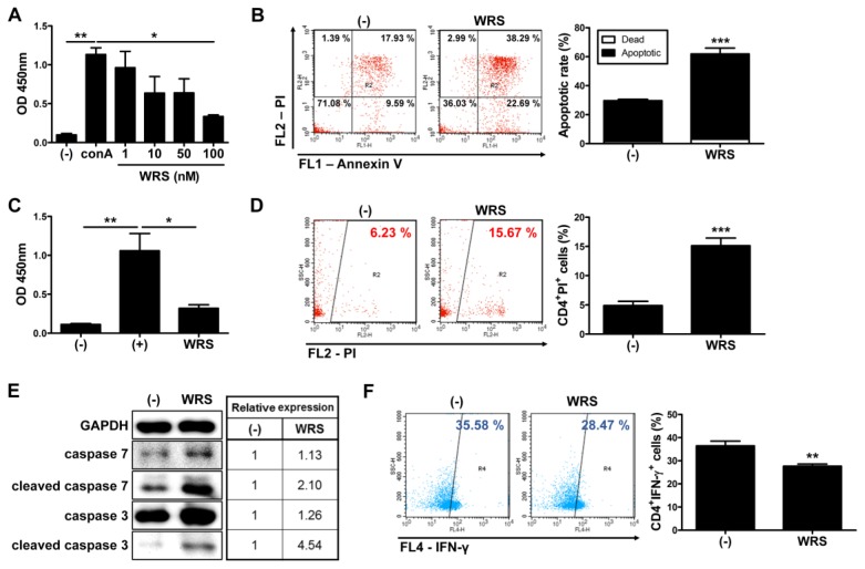
Fig. 3.
Secreted WRS inhibits the proliferation of CD4+ T cells by inducing apoptosis. Mononuclear cells and CD4+ T cells were isolated from human cord blood samples and cultured in the presence of WRS for three days. (A) Proliferation of hUCB-MNCs was measured by BrdU ELISA assay. (B) Apoptosis of hUCB-MNCs was determined by using flow cytometer. (C) Proliferation and (D) apoptosis of isolated CD4+ T cells were assessed. CD4+ T cells were isolated from hUCB-MNCs and cultured in the presence of WRS for three days. (E) Expression of signaling pathway molecules on apoptosis was determined by Western blot analysis. (F) Proportion of Th1 cells was measured by flow cytometric analysis after staining with cell surface CD4 and intracellular IFN-γ. *P < 0.05, **P < 0.01, ***P < 0.001. Results are shown as the mean ± SEM.

