Fig. 1.
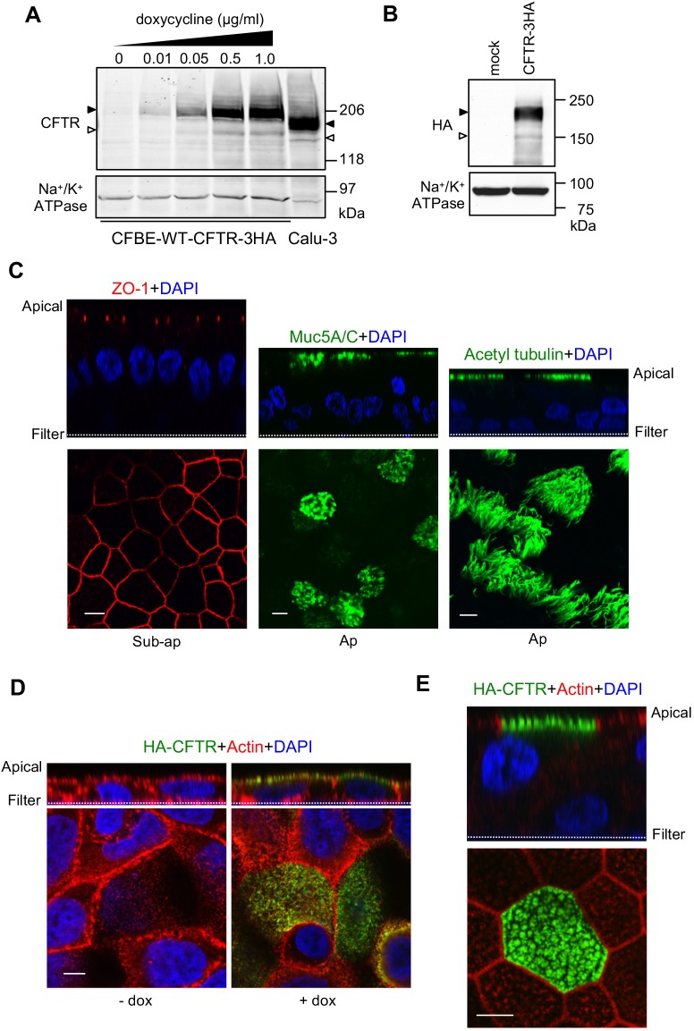
Characterization of human CF bronchial epithelial models to study polarized CFTR sorting. (A,B) Immunoblot analysis of CFTR–3HA expression (A) in CFBE cells after 3 days induction with the indicated amount of doxycycline and (B) following lentivirus transduction of CR-HBE cells. For comparison, an equal amount of Calu-3 cell lysate was loaded (A). Black and white arrowheads indicate complex- and core-glycosylated CFTR, respectively. Na+/K+ ATPase was probed as a loading control. (C) CR-HBE cells were differentiated at an air–liquid interface (ALI) and stained for tight junctions (ZO-1, red), mucin 5 (muc5A/C, green), cilia (acetylated-tubulin, green) and nuclei (DAPI, blue). Cells were visualized by laser confocal fluorescence microscopy. The upper and lower panels are vertical and horizontal optical sections, respectively. Ap, apical surface. (D,E) WT-CFTR–3HA is apically expressed in CFBE (D) and CR-HBE (E) cells. Cells were stained for WT-CFTR–3HA (green), actin (red) and DAPI (blue). Scale bars: 5 µm. Representative of at least three independent experiments.

