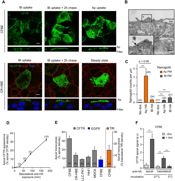
Fig. 2.
CFTR is apically transcytosed in human airway epithelial cells. (A) CFTR–3HA was labeled by anti-HA Ab capture (3 h, 37°C) from the basolateral (Bl) or the apical (Ap) compartment in filter-grown CFBE or CR-HBE cells. Cells were chased for 2 h (37°C) in the absence of anti-HA Ab. CFTR–anti-HA Ab complexes were visualized with AF488-anti-mouse Ab and laser confocal fluorescence microscopy. Lower panels represent z-sections. Scale bars: 5 µm. (B,C) Monitoring CFTR transcytosis with immuno-EM. (B) Immuno-EM detection of CFTR-3HA at the apical PM after basolateral labeling with anti-HA (3 h, 37°C) in CFBE. CFTR-anti-HA complexes labeled with secondary Ab conjugated to nanogold (arrow). The inset represents a higher magnification of the indicated area. Scale bar: 0.5 µm. (C) CFTR-expressing or non-expressing (no dox) CFBE cells were labeled either by apical (Ap) or basolateral (Bl) anti-HA Ab capture for 3 h (37°C). CFTR–anti-HA antibody complexes were visualized with anti-mouse Ab conjugated to nanogold particles as in B. The number of nanogold particles per micrometer was counted along the apical and basolateral PM segments (n=2–3 independent experiments, Mann–Whitney U-test). (D) CFTR apical transcytosis was measured over time as in Fig. S2B. (E) Apical transcytosis of CFTR–3HA, and endogenous EGFR and TfR were measured as described in the Materials and Methods in the indicated cells. (F) Apical transcytosis of CFTR is abolished when the channel is basolaterally labeled by anti-HA Ab for 3 h on ice or when CFTR transcription is not induced (- dox) (n=4). Data are means±s.e.m. on each panel. **P<0.01, parentheses indicate the number of independent experiments (D,E) or regions of interest (C).

