
Fig. 1.
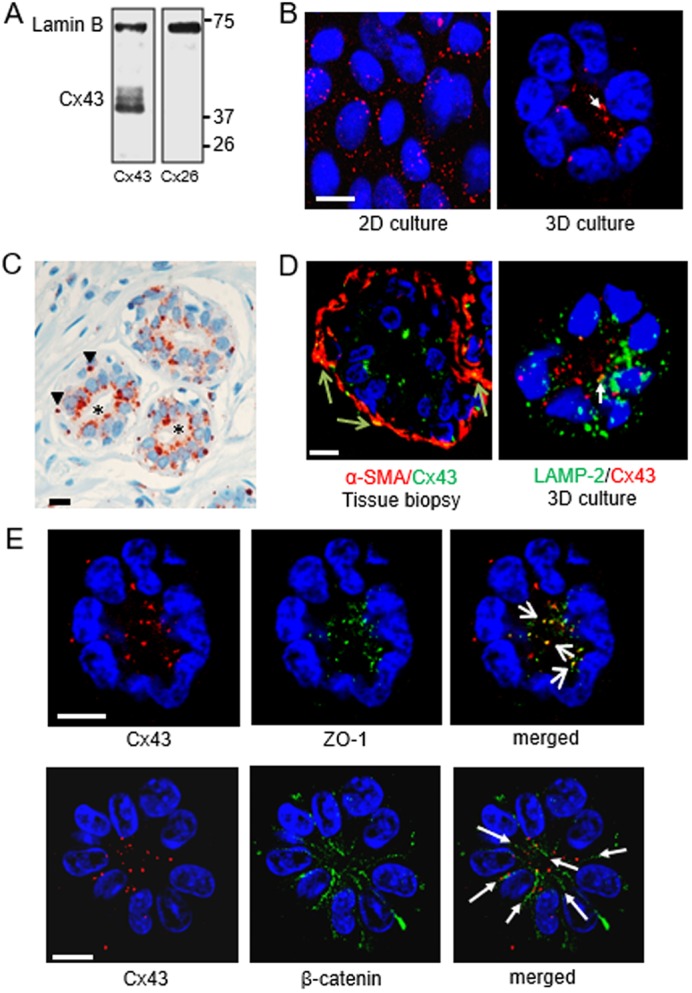
Cx43 is located apically in the breast luminal epithelium. S1 non-neoplastic mammary epithelial cells were cultured in 2D (A,B) or in 3D (B,D,E), as indicated, for 10 days. A thin section from breast tissue biopsy was used in C. (A) Western blot shows that Cx43, but not Cx26, is expressed in S1 cells; lamin B is used as loading control. (B) Immunostaining for Cx43 (red), with apical localization indicated by the arrow. (C) Immunohistochemistry for Cx43 (reddish-brown) in normal-appearing breast glandular tissue, with display of basal localization in myoepithelial cells (arrowheads) and apical localization in luminal cells (asterisks). Nuclei are counterstained with hematoxylin (blue). (D) Left: dual fluorescence staining for Cx43 (green) and a myoepithelial cell marker (α-smooth muscle actin protein, α-SMA; red) in normal-appearing breast glandular tissue. Cx43 staining overlap with α-SMA staining in myoepithelial cells appears in yellow (arrows). Right: dual immunostaining for Cx43 (red) and a lysosomal marker (lysosomal-associated membrane protein 2, LAMP-2) (green) in an acinus formed by S1 cells; the arrow points to a rare spot with colocalization (yellow). (E) Dual staining for Cx43 (red) and ZO-1 (green) or β-catenin (green). Colocalization of Cx43 and ZO-1 staining appears yellow (short arrows); cell–cell contacts with Cx43 aligned with β-catenin are indicated (long arrows). Nuclei are counterstained with DAPI (blue). Scale bars: 10 µm. Single immunofluorescence staining was done on multiple (>5) biological replicates (cell cultures and tissue samples); dual immunostaining was done on 2–3 biological replicates.