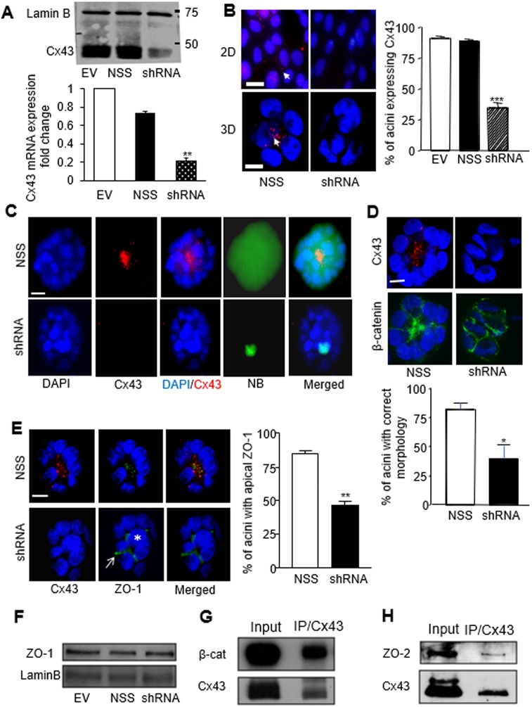
Fig. 3.
Downregulation of Cx43 expression disrupts GJIC and apical polarity in S1 cells. (A) Western blot (upper image) and quantitative real-time PCR (bottom graph) for Cx43 expression in S1 cells following retroviral delivery of empty vector (EV) or non-specific sequence shRNA (NSS) used as negative controls, and Cx43-specific shRNA (shRNA). Lamin B serves as loading control; n=3. Cx43 mRNA expression normalized to EV control; data represented as mean±s.e.m. (B) Fluorescence immunostaining for Cx43 (red) in S1 cells treated as indicated and cultured for 10 days in 2D or 3D conditions. Arrows indicate Cx43 foci. Graph shows the mean±s.e.m. percentages of acini that express Cx43 in each treatment condition; at least 200 acini were analyzed per condition; n=3. (C) S1 cells infected with NSS (upper panel) or Cx43 shRNA (lower panel) were cultured in 3D for 10 days and microinjected with 3% NB in 0.15 M LiCl, followed by dual fluorescence staining with streptavidin–FITC and an antibody against Cx43 (red). Merged images show the extent of NB spread within the acini; n=10 acini. (D) Dual immunostaining of acini for Cx43 (red) and β-catenin (green; indicating cell–cell limits) in S1 cells treated as indicated. The peripheral organization of cells around a hollow center (left lower panel) is considered morphologically correct. Graph shows mean±s.e.m. percentages of acini with correct morphology; at least 100 acini analyzed per condition; n=3. (E) Dual immunostaining for Cx43 (red) and ZO-1 (green) in acini formed by S1 cells treated as indicated. The arrow points to the peripheral location of ZO-1 and the asterisk indicates the central location of a cell (optical section through the middle of the acinus), illustrating abnormal morphogenesis. Graph shows mean±s.e.m. percentages of acini with apical ZO-1 staining; at least 100 acini analyzed per condition; n=3. (F) Western blots show unchanged levels of total ZO-1 expression in 10-day-old S1 acini treated as indicated. Lamin B was used as loading control. (G,H) Western blots for Cx43 and co-immunoprecipitated β-catenin (G) or ZO-2 (H) following immunoprecipitation with Cx43 antibody in S1 acini. *P<0.05, **P<0.01, ***P<0.001; one way ANOVA, with Dunn's comparison (A,B), nonpaired t-test (D,E). Nuclei are counterstained with DAPI (blue). Scale bar: 10 µm.

