
Fig. 4.
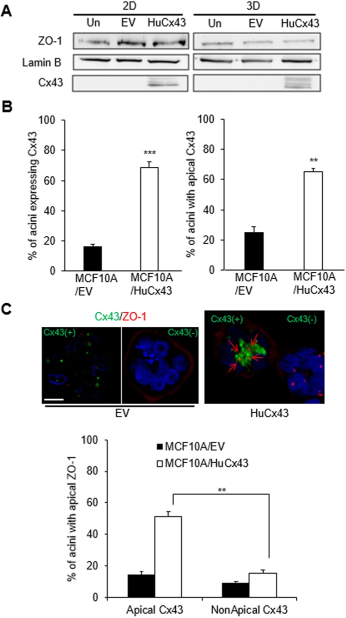
Cx43 expression induces apical polarity in MCF10A acini. (A) Western blots for Cx43 and ZO-1 expression from 10-day 2D and 3D cultures of MCF10A cells that were uninfected (Un) or infected with empty vector (EV) for controls, or infected with HuCx43 vector (HuCx43). Lamin B is used as loading control. (B) MCF10A cells stably infected with EV (MCF10A/EV) or with HuCx43 (MCF10A/HuCx43) were cultured in 3D for 10 days to induce acinar differentiation. Graphs show mean±s.e.m. percentages of acinar structures expressing Cx43 (left) and of acinar structures with Cx43 apically localized among those expressing Cx43 (right). (C) Representative images of dual immunostaining for Cx43 (green) and ZO-1 (red) in acini formed by MCF10A/HuCx43 and by MCF10A/EV with (Cx43+) and without (Cx43−) Cx43 expression. Red arrows on merged image indicate the apical location of ZO-1. Nuclei are counterstained with DAPI (blue). Graph shows mean±s.e.m. percentages of acini with apically localized ZO-1 among those with apical Cx43 and non-apical Cx43. A minimum of 100 acini were analyzed in each condition although the number of acini analyzed was less in EV population due to the paucity of structures expressing ZO-1 in C; n=3; unpaired-test (B) and one-way ANOVA with Dunn's comparison (C), **P<0.01, ***P<0.001 (significance only shown for comparison of interest in C). Scale bar: 10 µm.