Fig. 5.
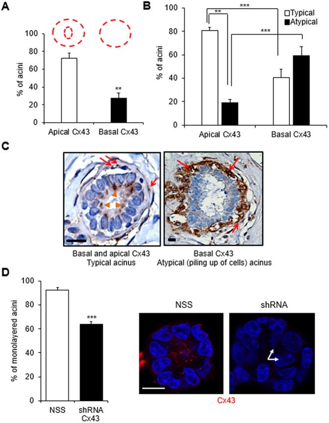
Absence of luminal expression of Cx43 is associated with cell multilayering. (A–C) Immunohistochemical staining for Cx43 was performed on archival breast tissue biopsy sections from 22 women with no history of breast cancer. Graphs show mean±s.e.m. percentages of acini with apical localization of Cx43 in the luminal epithelium (in addition to basal myoepithelial localization; see drawing in red) and basal localization of Cx43 only (A); percentages of acini displaying typical (one layer of luminal cells at the inner side of the layer of myoepithelial cells) or atypical (piling up of cells) organization depending on the location of Cx43 (B), n=22 acini; paired t-test. (C) Representative images of acini with basal (red arrows) and apical (orange arrowheads) Cx43 localization in a typical (normal-appearing) epithelial structure and only basal Cx43 (red arrows) in an atypical structure. Nuclei are stained with hematoxylin (blue). (D) S1 cells stably silenced for Cx43 expression were cultured in 3D for 10 days and immunolabeled for Cx43. Graph shows mean±s.e.m. percentages of acini with a monolayer of cells in acini of cells transduced with non-specific sequence (NSS) control and Cx43 shRNA. Shown are representative images of a monolayered acinus with Cx43 (red) apically localized and a multilayered (arrows) acinus lacking Cx43 staining. Nuclei are stained with DAPI. At least 100 acini analyzed; n=3, unpaired t-test. **P<0.01, ***P<0.001. Scale bars: 10 µm.

