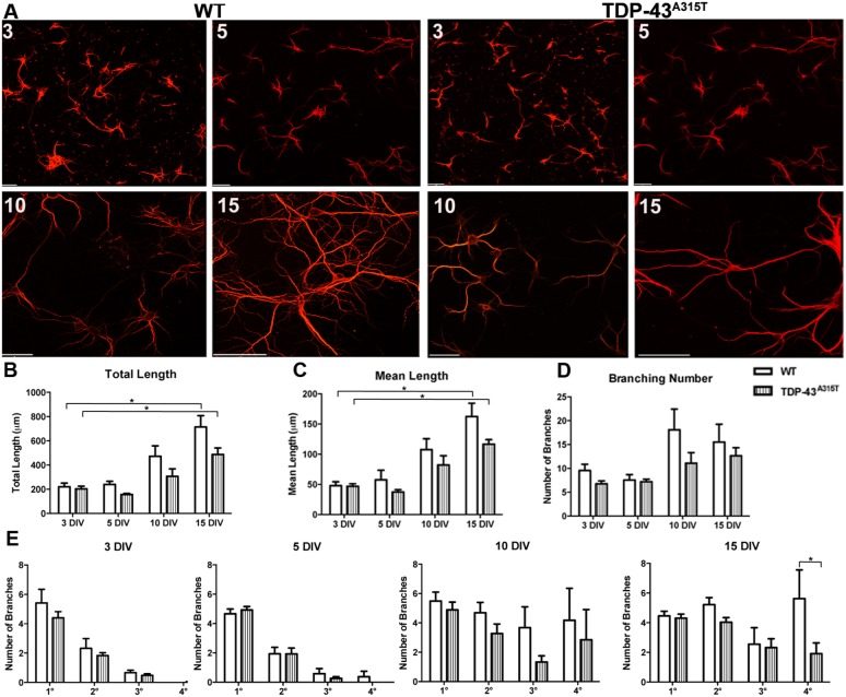
Fig. 2.
The effect of TDP-43A315T expression on dendrite development. (A) Primary cortical neurons derived from YFP:WT (left) and YFP:TDP-43A315T (right) single embryo cultures labeled for MAP2 at 3 DIV, 5 DIV, 10 DIV and 15 DIV. (B) Total dendrite length significantly increased between 3 and 15 DIV in both WT and TDP-43A315T cultures. There was no significant difference in total dendrite length between WT and TDP-43A315T at any time point. (C) Mean dendrite length significantly increased between 3 and 15 DIV in both WT and TDP-43A315T cultures. There was no significant difference in mean dendrite length between WT and TDP-43A315T at any time point. (D) There was no significant difference in dendritic branching number between 3 and 15 DIV in both WT and TDP-43A315T cultures. There was no significant difference in dendritic branching number between WT and TDP-43A315T neurons at any time point. (E) Quantification of the number of primary (1°) secondary (2°) tertiary (3°) and quaternary (4°) dendrite branches at 3, 5, 10 and 15 DIV demonstrated that TDP-43A315T neurons became significantly less complex in quaternary branches by 15 DIV. n=3 biological replicates. *P<0.05 (two-way ANOVA with Tukey's multiple comparisons test). Data are mean±s.e.m. Scale bars: 20 μm.

