Fig. 4.
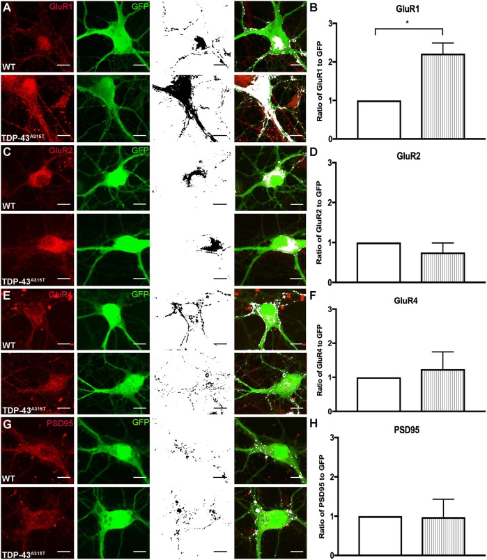
The effect of TDP-43A315T expression on synaptic receptor expression. (A-H) YFP:WT and YFP:TDP-43A315T primary cortical neurons at 10 DIV were labeled for postsynaptic receptors and GFP (antibody against GFP enhances endogenous YFP fluorescence). Immunocytochemistry labeling for the glutamate receptor subunits (red, first column) GluR1 (A), GluR2 (C) and GluR4 (E) and the postsynaptic protein PSD-95 antibody (G), and GFP antibody (green, second column); colocalization (black, third column) was identified in ImageJ and overlaid onto GFP fluorescence images (white; fourth column). (B,D,F) The ratio of GluR1 to GFP was significantly increased (B), whereas there was no significant differences in the ratio of GluR2 to GFP (D) and GluR4 to GFP (F) in the TDP-43A315T neurons in comparison to WT controls. (H) There was no significant difference in the ratio of PSD-95 to GFP in the TDP-43A315T neurons in comparison to WT controls. n=3 biological replicates. *P<0.05 (Student's t-test). Data are mean±s.e.m. Scale bar: 10 μm.

