
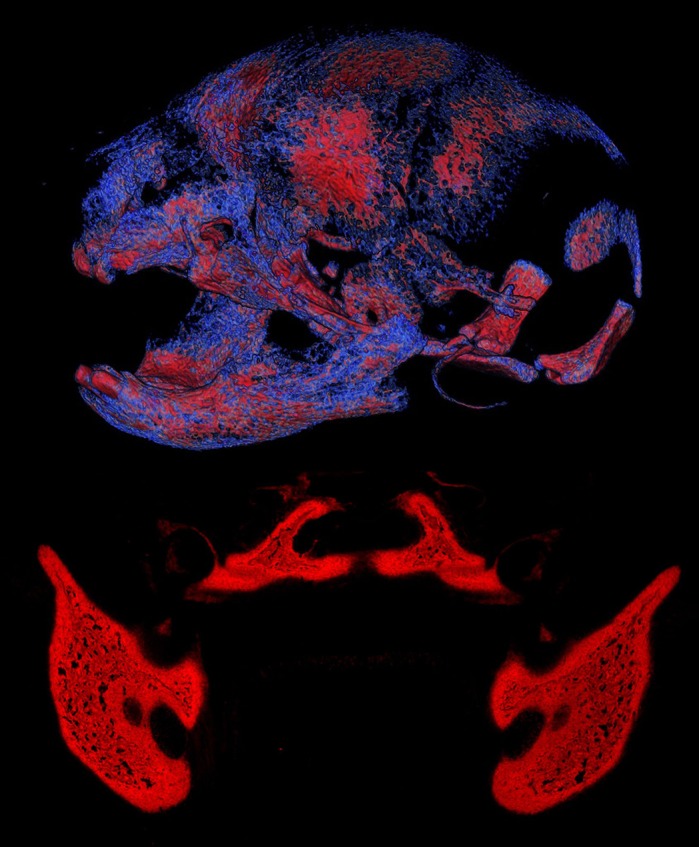
Craniofacial tissues of an Apert syndrome mouse and wild-type embryo. Top: volume rendering of mineralized craniofacial tissues (lateral view of skull) from µCT images of a newborn (P0) Fgfr2+/S252W Apert syndrome mouse (image by S.M.M.P.). Bottom: the osteogenic tissue in the mandibular and palatine regions of a wild-type mouse embryo at E16.5, visualized using alkaline phosphatase staining (red) (image by M.W.).