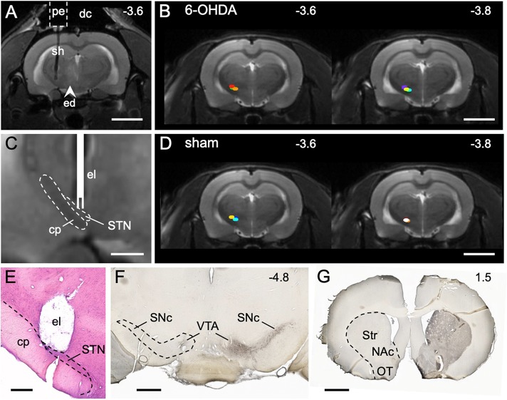
Fig. 1.
STN stimulation site and dopaminergic lesion. (A) MRI (T2) 24 h after implantation of the guide cannula and 6-OHDA injection. The pedestal (pe) of the guide cannula is attached to the skull with dental cement (dc). The shaft (sh) targets the STN. A faint edema (ed) is visible from 6-OHDA injection into the medial forebrain bundle. (B) Stimulation sites (colored dots) of 6-OHDA animals (n=7). (C) MRI detail from the stimulation site with sketched electrode (el). The cerebral peduncle (cp), which contains descending motor fibers, lies next to the STN and should not be stimulated. (D) Stimulation sites (colored dots) of sham animals (n=6). (E) Histological section showing the stimulation site after removal of the electrode. (F) TH immunostaining (transverse section level) demonstrating the loss of dopaminergic cell bodies in the left substantia nigra pars compacta (SNc) and ventral tegmental area (VTA) in a rat injected with 6-OHDA. (G) TH immunostaining (transverse section level) showing the loss of dopaminergic axon terminals in the left striatum (Str), nucleus accumbens (NAc) and olfactory tubercle (OT). Numbers are rostrocaudal coordinates (mm) relative to Bregma. Dashed outlines indicate borders of brain regions. Scale bars: 5 mm in A,B,D; 1 mm in C,F,G; 500 µm in E.

