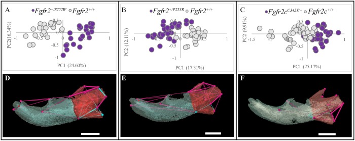
Fig. 1.
Morphological differences in newborn (P0) mice carrying mutations associated with three FGFR2-related craniosynostosis syndromes and their unaffected littermates. (A-F) Results of PCA of mandibles based on unique linear distances among 3D landmarks (A-C) and EDMA of landmark coordinates (D-F). Scatter plots of individual scores on first and second PC axes (PC1 and PC2) of linear distance-based PCAs of the hemimandibles of mutant and unaffected littermates of Fgfr2+/S252W and Fgfr2+/P253R Apert syndrome mouse models (A,B, respectively) and Fgfr2cC342Y/+ Crouzon/Pfeiffer syndrome mouse model (C). Results of EDMA of each craniosynostosis mouse model and unaffected littermates showing linear distances within each model that are significantly different by at least 5% between mutant and unaffected littermates (D-F). Blue lines are significantly larger in mutant mice relative to unaffected littermates; fuchsia lines are significantly smaller in mutant mice. Thin lines indicate linear distances that are increased/decreased by 5-10% in mice carrying one of the Fgfr2 mutations whereas thick lines indicate linear distances that differ by >10% between mutant and unaffected mice. The buccal aspects of the left hemimandibles of the models were used for illustration. Hemimandibles were segmented into an anterior portion (anterior body, blue) and posterior portion (ramus, red) to indicate functional areas. Scale bars: 1 mm.

