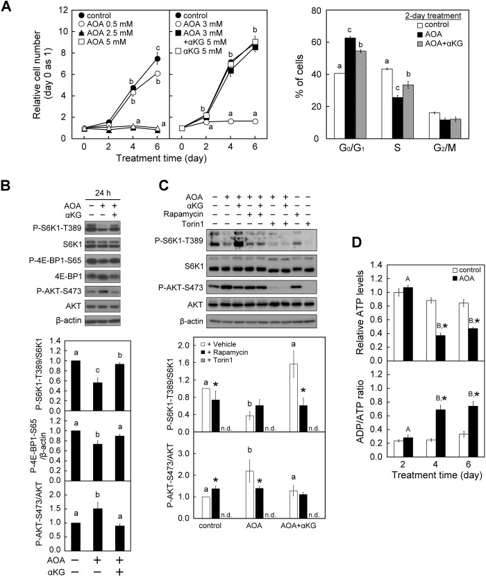
Fig. 1.
Inhibition of glutamine-dependent anaplerosis with AOA induces inhibition of cell proliferation and cell cycle arrest in WI38 cells. WI38 cells were treated with vehicle or AOA in the absence or presence of 5 mM αKG for the indicated time-period with medium changed at 2-day intervals. (A) Effect of AOA on cell proliferation and cell cycle progression. Left panel: the relative cell numbers were calculated by normalizing against the value of day 0. Right panel: cell cycle analysis was conducted after a 2-day treatment period using flow cytometry with Propidium Iodide staining of DNA. The percentages of cells in various phases of the cell cycle are presented. (B) Effect of AOA on mTORC signaling. Cell lysates were prepared after a 1-day treatment period and analyzed for the activation status of mTORC1 and mTORC2 by immunoblotting and densitometry analysis of P-S6K1-T389/S6K1 and P-4E-BP1-S65/β-actin, and P-AKT-S473/AKT with β-actin served as a loading control. (C) Effect of mTOR inhibitors on AOA-regulated mTORC1/2 activities. Cells were first treated with 3 mM AOA and then given vehicle (DMSO), 1 nM rapamycin or 30 nM Torin1 1 h before the end of the 24-h culture period. Cell lysates were analyzed by immunoblotting as described in B. (D) Effects of AOA on energy status. Intracellular ATP content and ADP/ATP ratio were determined by ApoGlow Assay Kit. All quantitative data are expressed as the mean±s.e.m. (n=3) of three independent experiments. Different lowercase letters indicate significant difference among treatment groups at the same time-point (P<0.05). Asterisk (*) designates a significant difference compared with the respective vehicle control (P<0.05).

