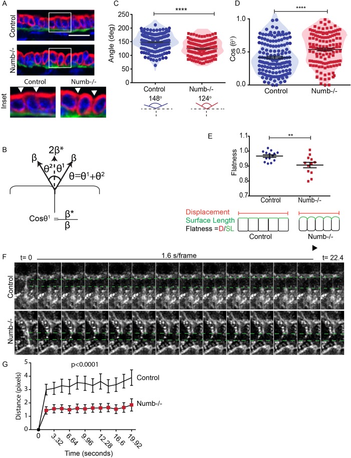
Fig. 8.
Numb regulates cell tension in mammary epithelial cells. (A) Fluorescence of images of control and Numb-deficient mammary glands immunostained for CK8 (red). White arrowheads indicate points of cell–cell contact. (B) Diagram showing measured angles between adjacent cells: β, cortical tension at the free surface relates to β*, the tension at cell–cell contacts, through cosine of the angle between them. (C) Scatter plot of θ between adjacent control or Numb-deficient cells. n=140–179 neighboring cells. (D) Scatter plot of Cos(θ1) for control and Numb-deficient cells. n=130–147 cells. (E) Scatter plot of the epithelial duct flatness along the apical surface. 12–14 ducts from both groups. (F) Time-lapse images of a control and Numb-deficient before (t=0) and after laser ablation. Green dashed lines indicated the deviation of the membranes from the t=0 position after ablation. (G) Quantification of the recoil distance following laser cutting of mammary epithelial cells (P<0.0001). n=25–27 cells from both groups (paired t-test, two-tailed). Error bars=s.e.m. Unpaired t-test; two-tailed: ****P<0.0001, **P<0.01. Scale bar: 20 μm.

